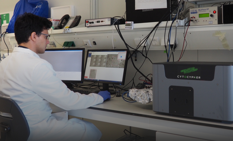
Q&A Report: A Comprehensive How-To Demonstration of Higher Throughput Excitation-Contraction Coupling Investigations

How easy is it to detect an arrhythmic event? Can calcium dyes interfere with detection of arrhythmic events?
M. Helmes: Arrhythmic events are easiest to detect in the contractility tracing, so you wouldn’t have interference from calcium dyes. The effect of calcium dyes is twofold. It will always buffer calcium to some extent, which could possibly interfere with arrhythmic events. The other is that the dyes always have some modest toxic effect, possibly enhancing arrhythmias. My inkling would be to use contractility to detect arrhythmic events (reference Abi Gerges), and use calcium dyes to help figure out what has changed in the calcium handling to explain the arrhythmias.
Does the computer remember the cells studied and the order of study?
M. Helmes: It does. The location from each measurement is stored and can be returned to. Within the same file it will indicate whether it is the first, second, etc. measurement. The list can also be recalled in a new file so you can revisit the same cells.
How fast can you change the solution in the chamber?
M. Helmes: Depends on the method of exchange. Using pipetting it can be done in about half a minute. With perfusion you probably need minutes to have sufficient exchange. We originally though you couldn’t use pipetting and re-measure the same cells, but in practice, if you are careful, it turns out that you can. It is of course riskier, more chance to wash away cells or bumping dish and thereby offsetting the position of the cells.
Has this device ever been used to address smooth muscle cell contraction?
M. Helmes: Not that I am aware of. I don’t think it is fundamentally different compared to amorphous ips-derived cardiomyocytes, whose contraction we can measure. Would love to try it though.
We are currently struggling with Rat isolation, experiencing huge variation and concerning viability. Any suggestions?
M. Helmes: This is difficult to answer in one or two sentences as it can have many causes. Consistent cannulation is of course the first important step. Using a filtered solution so you have no small particles blocking the blood vessels is also important. Using a very consistent temperature of perfusate of the heart so you get consistent digestion times is essential. Ensure you have clean tubing so there is no bacterial growth. You have to replace the tubing occasionally and definitely if you have problems with the isolation.
Is it possible to determine excitability of skeletal muscle fibers with the system?
M. Helmes: We have determined the contractile properties of FDB myocytes on the system using twitches. We tested the effect of CK, a calcium sensitizer, on contractility in repeated measures and got consistent results.
Is this system and the automatic analysis compatible with general fluorophores... i.e. to distinguish between GFP positive cells and negative cells within a dish?
M. Helmes: It is not something we have tried but here is how I think it could be done using photometry. Measure fluorescence with the first run, using the PMT. Take the average fluorescence signal. Possibly normalize it to the size of the cell (an image of each cell can be saved) and plot the parameters of interest against the fluorescence of the cell. We do not as of now have to possibility to select cells based on fluorescence levels, although this wouldn’t be too hard to implement in case you’d want it.
Can cardiac contractility/calcium handling be measured in 3D cardiac organoids?
M. Helmes: I would think so, provided you can visualize them on the microscope, as it is a general contractility/fluorescence system. It would of course only make sense if you can put a large number of 3D constructs in the system, like in a 24 well plate. We have algorithms in the software that can measure the deviation of for example a PDMS pillar to which the 3D organoid is attached.
How does the system change excitation wave length when using a ratio-metric dye?
M. Helmes: We drive two different wavelength LEDs alternatively and de-multiplex the signal from the photo-multiplier tube that measures the emission light.
Can the automatic analysis also analyze sodium dye?
M. Helmes: It depends on the shape of the signal. The assumption in the analysis is that there is a transient departure from baseline. We currently don’t support just taking the average from a trace, but that would be an easy adjustment in the analysis software that we would be happy to make for you.
This is not related to myocytes, but I am currently using skeletal muscle cells and investigating contractility for secretome study. Is there a similar software for detecting calcium in muscle cell using this device?
M. Helmes: We have used this system to measure skeletal myocytes (FDB myocytes). This can be done. Please contact us if you want detailed information on this. We can share the data with you.
Can the system measure AP and CaiT simultaneously?
M. Helmes: Yes, it can, as can be seen in the webinar where we measure AP, Ca++ and sarcomere length simultaneously.
Can you measure amplitude of contraction in iPSCMs with the multicellular setup?
M. Helmes: No, you cannot. At least not with the algorithm shown. We use the correlation between a reference frame and the current frame to assess the relative change in shape (contractility). Absolute length cannot be deduced from a correlation coefficient. This means you cannot use percentage shortening as you may be used to. We have another algorithm that will allow you to do this (EdgeBox), but it is more prone to artifacts and harder to align, so you will have to sacrifice throughput.
Can one perfuse the cells and switch among perfusates using this system?
M. Helmes: Yes, you can. With the pump system that can be delivered with this system you can switch between up to 6 different solutions in one experiment.
If you measure the contraction parameters in an iPS monolayer, how do you know how many cells are included?
M. Helmes: You cannot know this as there is no way (at least with this system) to distinguish individual cells within a monolayer. As the cells within the region of interest are all coupled, we think that our measurement gives a realistic assessment of the contractility of that population of cells.
Could calcium of non-contractile cells be measured as well?
M. Helmes: In principle yes, as it measures (ratiometric) calcium. But you do have to have a way to identify the cells you want to measure. Keep in mind that as of now it is not an imaging system but a photometry system, that will give you no spatial information except that you know the select circle that is illuminated.
How reproducible are the effects you see on the screen (as you only measure compounds twice)?
D. Kuster: It is quite reproducible. Our current approach is to measure every compound twice. Compounds that show an effect in either of the measurements days (or both) are measured a third time. Most compounds show a reproducible effect size on different measuring days. You have to check control values of the individual days though. And randomizing of the compounds is essential.
You use kinase inhibitors in your screen, but most of the compounds increase contraction. Is that what you expected?
D. Kuster: Not really what we expected, but it might be due to the fact that the cells are not very strongly stimulated to begin with. This might make it easier to pick up activators than inhibitors of contraction. But it is clear that a lot of kinases inhibit contraction, which is an interesting finding.
Why did you select TTB 70%, did the other TTB values have the same significance? Why not use tau from monoexponential fit, or biexponential data?
D. Kuster: This is a good question. Other time to baseline values showed the same effect (albeit with not exactly the same effect size). I always like 70% because it describes a sizeable amount of the relaxation, but is does not suffer from problems with fitting the return curve (sometimes if the contraction is small, the 90% baseline is more difficult to measure correctly). We also take along tau to see if the same changes are also seen there. I think the combination of return velocity and either time to baseline or tau tells you most of what you need to know about relaxation.
Were your measurements automatic or by hand?
D. Kuster: We did that particular data set completely manually. But this is now completely based on mouse clicks and keyboards commands. So, thankfully, no more rotating of the camera to get the cell straight. For newer projects we do either manual or automatic depending on which cells we use (if the yield is good, i.e. healthy rats, automatic is better).
What is the final calcium concentration of cells?
D. Kuster: We use 1 mM of Ca2+. I prefer this over 1.8 mM that we used previously and that is often seen in literature. The cells seem to be more stable in 1 mM, especially when you are testing an inotropic compound such as iso.
Why did you only do 15 min/well?
D: Kuster This had more practical reasons as we wanted to increase throughput and within 15 minutes, we would always be able to measure 30 to 40 cells. We have checked if run-down occurred during the 15 minutes and found no time dependent effect. However, we have measured cells for longer (including repeated measures) and saw no consistent effect. But this only holds true when cell quality is good to begin with.
You measured more than 1000 cells and only included 700 cells, what was your exclusion criteria?
D. Kuster: We try to measure every cell that moves to not create a bias towards the best-looking cells. The main reason for excluding cells is that we are not able to get good transients (with good being defined as having a good fit). The exclusion criteria are published in our paper in Frontiers in Physiology 2020 paper (Nollet et al.) and are based on the number of contractions and the goodness of fit. These characteristics were based on rat cardiomyocytes, with mouse cardiomyocytes we are a little bit more lenient (albeit still quite stringent) on the fit parameters.
What parameters do you use when you screen hundreds of cells to select healthy versus unhealthy/damaged cells?
D. Kuster: I am primarily interested in relaxation, so I like to use a combination of time to 70% baseline (as this describes most of the relaxation phase and yields reproducible results), return velocity and tau.