Q&A Report: Hippocampal Neural Circuits in Awake Head-Fixed Mice Navigating a Real-World Environment
How do you define place cells? Do you work with deconvolved or raw fluorescence?
Simon Schultz: See the Methods section in the Frontiers paper for full details. Briefly: we use deconvolved traces then detect events, and keep an “event train” where each even has an amplitude (note difference to spike train). For the circular track, we have 3 criteria: (i) events present in at least half of laps, (ii) events present for > a fixed threshold of time bins when mouse was moving within the place field (threshold obviously depends on bin width), (iii) Skaggs spatial information exceeding 99% of values for shuffled data. Then there is a related procedure for the full 2D place fields.
Can we conduct similar experiments in rats?
Neurotar: The Mobile HomeCage can be used with neonatal and juvenile rats whose weight is not exceeding 75g. There are no weight restrictions for mice.
The implant causes some damage to the cortex and hippocampus. Does the physical damage resulting in some visible and significant behavioral or memory impairments?
Michael Goard: There are no overt behavioral or memory impairments, though we have not run thorough tests that may reveal more subtle deficits.
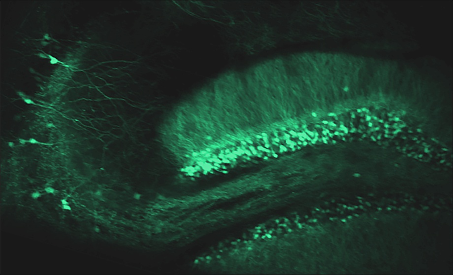
Are the shifting place fields over time completely random, or is there a correlation between different cells that have a more stable relative relationship?
Simon Schultz: Not random. They seem to drift and jump, in some cases being metastable between two locations. See Fig 5A of the Frontiers Cellular Neurosci paper. I’m not sure a correlation coefficient would capture what is going on, but I think you might get it out of some of the attractor models etc.
I'm interested in using the system to study spine dynamics without the confounding factors of anesthesia. Could the awake state of the mouse (i.e. movement) act as a confounding factor?
Neurotar: The Mobile HomeCage was tested for spine dynamics imaging in awake mice (see Pryazhnikov et al., 2019). Mouse movement increases image displacement to some extent, but the imaging stability is sufficient for reliable data analysis even during the running state.
Mark Schnitzer’s lab showed very high turnover of spines in CA1. I am wondering how your data compares with their data?
Michael Goard: We also see high turnover rates compared to the neocortex. The Schnitzer lab focused on basal dendrites since they can be seen from the upper surface of the hippocampus. We are in the process of quantifying spine turnover rates in CA1 apical dendrites to see how they compare.
Do some cells that were not place cells become PCs on subsequent days or environments?
Simon Schultz: Yes. And vice versa.
What resolution are you able to achieve?
Neurotar: The resolution parameters are determined by a method that is used for imaging. For example, in two-photon imaging, one can detect dendritic spines but not the synaptic proteins at the molecular level.
What is the working distance of the objectives used when imaging with hippocampal microprisms?
Michael Goard: We used a Nikon 16x/0.8NA objective with a 3mm working distance. Since we were imaging through 2.0 – 2.5mm of glass, we were close to the limit of the working distance of the objective, so it was important that the window was completely parallel to the objective and that there was no dental cement above the window that could limit movement (or even scratch the objective surface).
Why is the light dimmed over the session, and why is it lit in the first place and not just tactile stim?
Simon Schultz: That is because of the physics of phosphorescence. And it is lit to provide a more full sensory representation of the environment.
Is there a reward? How do you motivate the animals to keep running?
Neurotar: To motivate animals to run, they were given a water reward of 4 μL per loop traversed at random locations. Daily water intake was limited to 1–3 mL and was individually adjusted for each mouse to maintain the target weight of 85% of the pre-restriction weight. At the start of each training session, two rewards were given to motivate the mice to lick for water. There was no limit to the number of rewards animals could have during sessions.
Can spine density be affected by hormones in male mice?
Michael Goard: Yes, there is evidence that sex steroid hormones, such as testosterone (and estradiol, for that matter), affect spine density in male rodents.
Have you thought about including an LED screen that moves with the apparatus? This might let you understand the influence of cues without confounding stationary ones.
Simon Schultz: We thought about it – but very technically tricky, as the weight would need to be very very light to avoid changing the kinematics. Maybe with thin film technology, but then you still need to power it. I’m not sure this is viable.
Can you image hippocampal interneurons?
Michael Goard: We believe so. We are planning implants of Gad2-GCaMP6s mice in the near future.
I am doing in vivo hippocampal CA1 neuron calcium imaging with head fixed mice to see the basal activity of the neurons. I found the basal activity was higher on the first day when I put the mice in the Mobile HomeCage compared to the 3rd day when mice are more familiar with the environment. Is that normal?
Neurotar: This phenomenon can be observed if imaging was done soon after surgeries without allowing enough time for recovery. Usually, we advise doing imaging not earlier than three weeks after surgery. Alternatively, it could reflect the new environment exploration phenomenon (in case mice were not accustomed/trained in the cage prior to the imaging).