Q&A Report: Neurovascular Coupling: Novel Insights from Studies in Awake Head-Fixed and Anesthetized Mice
These answers were provided by:
Adrián Rodríguez-Contreras, PhD
Associate Professor
Communication Sciences and Disorders
Northwestern University
Barbara Lykke Lind, PhD
Assistant Professor
Department of Neuroscience
University of Copenhagen
Similarly to what is seen in astrocytic calcium, it appears the rises in calcium appear after the dilation. Can you comment on the timing of the events?
Barbara Lind: The astrocytic response occurs in two parts. One before and one after the dilation. That is true for PV-IN evoked responses, not the VGAT-IN dilations which were earlier.
Does this isoflurane inhalation protocol generate acidosis? GCaMP baseline fluorescence is sensitive to pH changes and can shift baseline fluorescence.
Adrián Rodríguez-Contreras: This is an interesting question that has two parts. The first part relates to the generation of acidosis when isoflurane is used. Isoflurane induced acidosis has been documented in animals that are generally anesthetized for periods of time that simulate the duration of surgical procedures (e.g., 60 minutes; Wilding et al., 2017, https://pubmed.ncbi.nlm.nih.gov/28315643/). It is important to note that mice in our study were exposed to isoflurane for up to 15 minutes (5 minutes at 5%, and 10 minutes at 1.5%). The second part relates to potential effects of acidosis on baseline fluorescence of GCaMP. The mice used in our study express GCamP8, which has been shown to have a lower baseline fluorescence compared to other GCamP variants (Ohkura et al., 2012 DOI: 10.1371/journal.pone.0051286). We do not know if acidosis effects have been characterized for the GCaMP8 variant.
The last study is of particular importance in the role of GABA in NVC as previous studies base this coupling in particular on glutamate activation of metabotropic receptors on astrocytes. What is your take on this?
Barbara Lind: In my view the effect of GABA in NVC appears to be comparable to the glutamatergic effect as the neurotransmitter works indirectly on the vascular diameter, by an astrocyte dependent mechanism. It is of course possible that it could be via the GABAergic metabotropic receptors, but that I cannot say yet. What I found was that astrocytic responsivity to GABA appears to depend on NA levels.
How deep do you image penetrating arterioles in PV optogenetic activation experiments?
Barbara Lind: All experiments were done in layer II/III of cortex; so from 100-250um below the surface.
Referring to Rungta's 2016 Nat Communications paper, was there any vasodilation to blue light illumination in control mice without Channelrhodopsin?
Barbara Lind: We did control experiments in animals not expressing Channelrhodopsin and found that our stimulation did not prompt vasodilation.
In the first study, could the initial Ca rise be as a result of photoactivation rather than (or combined with) sensory activation?
Barbara Lind: The responses we have are not to sensory activation but to activation of the PV interneurons. The response to the photoactivation should be the same in the PV and the VGAT mice (as we used similar stimulation intensity and duration), but we only see the first part of the astrocytic Ca2+ response during the PV stimulation.
Do you think the increase in intravascular viscosity because of Texas Red contributes to any changes in NVC?
Barbara Lind: It is possible that it could contribute non-physiologically. The only comfort in this consideration is that the dextran affected all experiments equally (same loading in the animals).
How long can you track calcium signals (minutes, hours, days)?
Barbara Lind: I track Ca2+ signals over minutes in the individual acquisitions, but I can return to the same FOV and follow the changes there over months (as long as my ethical permit allows).
In one of your slides, you showed that the processes of the astrocytes respond before the endfoot. I wondered if the response you see after noradrenaline administration (in which calcium response is reduced) is due to the location of different receptors along the astrocytes because you still see a minor calcium response. From where is this response coming?
Barbara Lind: It is a very interesting question, and I must confess that I have not thoroughly investigated the position of the remaining activity after removing the cortical NA levels. Based on a preliminary visual examination of my results I would say that it affects all areas equally or mostly the processes near PV interneurons.
How do you control for the movement of mice in the analysis?
Adrián Rodríguez-Contreras: That will depend on the purpose of the experiment. Tracking mouse movement simultaneously with imaging is now feasible with a mobile cage setup from Neurotar.
Barbara Lind: We can detect the movement onset and speed with the Neurotar cage. Based on this information we can classify and possibly disregard periods of movements.
Somatosensory stimulation may evoke emotional responses. I would like to know how experts distinguish cerebrovascular responses induced by sensory stimulation per se from those by emotions in awake animals.
Adrián Rodríguez-Contreras: This is an important question. However, I do not know of any reliable way to measure emotions physiologically. Some people are interested in measuring cardiac output or pupillary dilation as correlates, but I do not think those measurements can directly address the question asked.
Barbara Lind: It is true there is an overlap, and it can be difficult to determine how much the emotional response is due to sensory stimulation in an awake animal. Experiments can evaluate change in mouse movement or follow dilations of the pupil to detect for arousal during stimulations.
It has been suggested that sleep may regulate NVC. To what extent do you think the research using anesthetized mice may represent what happens during/after sleep?
Adrián Rodríguez-Contreras: Anesthesia can be seen as a pharmacologically induced brain state, which in my opinion is quite different from natural sleep. However, it may be worth considering comparative studies where physiological measurements are obtained, as they could illustrate similarities and important differences between chemically induced and natural brain states, e.g., electrical oscillations, memory processes. Exciting new research also indicates that activity in a glymphatic system is regulated in different ways during wake and sleep cycles, and that it is also affected by anesthetic regimes. See a recent review: https://pubmed.ncbi.nlm.nih.gov/30883420/
Any Ideal or recommended analysis tools/software?
Adrián Rodríguez-Contreras: Image J to start. Proficiency in computer coding and image processing tools will be needed as your studies progress. https://imagej.nih.gov/ij/index.html
Barbara Lind: I have been writing my own Matlab scripts to analyze, but now some good online opensource tools exist, such as aQua for astrocyte Ca2+ activity and GitHub for animal behavior analysis.
Astroglia participate in estrogen-induced synaptic plasticity. Astroglia respond to estradiol by decreasing glial process presence on neurons, allowing increased contacts/synapses on neurons, or the reverse in cases of decreased estrogen. Similar effects are seen on glia limitans. Please address hormonal effects in your talks.
Adrián Rodríguez-Contreras: In vivo experiments are always complicated by systemic variables. It is known that important hormonal differences exist between males and females. We used male and female mice in our studies. We did not observe obvious differences between the measurements obtained with respect to sex. To address the important role of hormones, one would have to manipulate them in a controlled fashion.
Barbara Lind: Hormonal responses in astrocytes are very interesting an unfortunately not something I have investigated in the context of GABAergic neurovascular coupling.
Would it be possible to address the question of NVC at the basis of functional connectivity, with the hypothesis of Mateo et al. (2019) and vasomotion, as well as other slow signal sources like Mayer's waves?
Barbara Lind: I believe it would be most relevant to do so.
Which anesthesia may interfere with my experiment?
Adrián Rodríguez-Contreras: Anesthesia induces an altered brain state. Different types of anesthetics will have different effects on physiological variables. The mechanisms of anesthetic action are not entirely understood. Research on anesthesia is relevant to improve and develop applications in medicine.
How do the different anesthetics affect the cerebral blood blow and brain metabolism, NVC and functional connectivity?
Adrián Rodríguez-Contreras: In different ways, depending on anesthetic type, age, and health state of subjects. For reviews:
https://pubmed.ncbi.nlm.nih.gov/?term=Effects+of+anesthesia+on+brain+review
https://pubmed.ncbi.nlm.nih.gov/28586779/
Barbara Lind: Most anesthetics are found to influence blood flow. Alpha-chloralose given continuously I.V. may be the least confounding.
What imaging setups did you use for the experiments?
Adrián Rodríguez-Contreras: Ultima in Vivo microscope from Bruker (Formerly managed by Prairie Technologies).
Do you see any movement artefact?
Barbara Lind: There can be problematic movements which will disturb the imaging precision and thus need to be excluded from the data. The tracker from Neurotar can give valuable information on what could be critical periods.
How long can you record in a single session, and how reproducible are the images if you acquire data at a later timepoint?
Barbara Lind: Our animal permit does not allow us to image for more than an hour in a single session, but reproducibility is quite possible if the experimenter is careful to find the exact same image FOV.
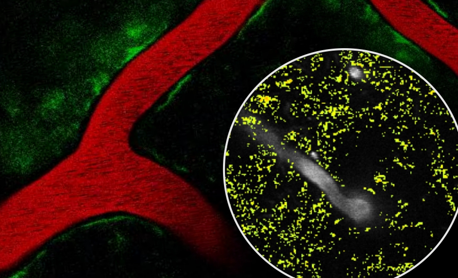
How was the astrocytic Ca analyzed, what about aQua?
Barbara Lind: We found that the identification of spatially and temporally correlated activities, an analysis partly similar to what is done with the aQua analysis tool, was very valuable to find the larger astrocytic Ca2+ activities. The method would on the other hand not efficiently pick up the immediate astrocytic response to the stimulation, possible because this occurred diffusely and primarily in the gliapil. So, the summation of the data was done using average of the FOV and for spatial investigation the Ca2+ fluorescence was aligned to the optogenetic stimulation.