Experts discuss how functional ultrasound imaging in awake head-fixed mice can help advance basic neuroscience and neurovascular research, as well as preclinical drug discovery.
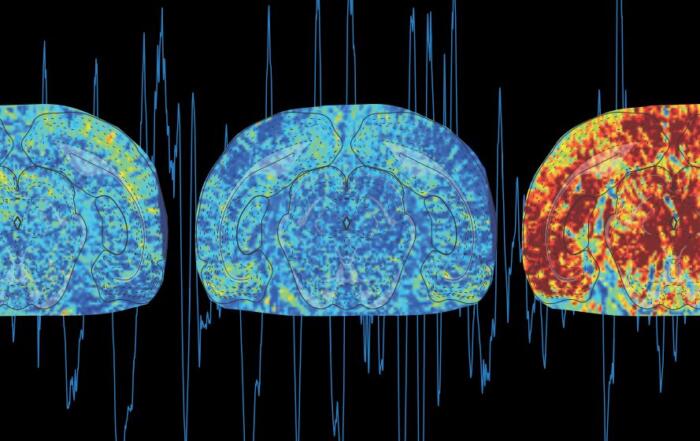
Functional ultrasound (fUS) imaging is a new kid on the block in neuroimaging. It combines high spaciotemporal resolution with deep tissue penetration, which enables non-invasive whole-brain imaging in mice.
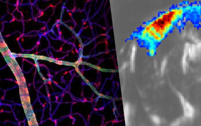
This exciting new technology complements and extends classical imaging modalities: it enables more straightforward, unobstructed and non-invasive functional measurements in mouse models of CNS diseases. Sensitive to changes in cerebral blood volume, fUS imaging is used to characterize brain networks with functional connectivity analysis and to measure the responses to sensory stimuli and pharmacological challenges.
fUS imaging performed in the brain of awake mice removes the biases and artifacts associated with the use of general anesthesia, which is no longer a “necessary evil” of translational imaging. Besides that: fUS imaging in awake mice allows integrating functional imaging with behavioral readouts starting from open field locomotion tracking to maze navigation and sociability studies.
Key Topics Include:
- Functional ultrasound (fUS) imaging methodology
- How translational fUS neuroimaging helps to advance basic neuroscience research and preclinical drug discovery
- The main advantages and limitations of using functional ultrasound compared to other techniques such as BOLD fMRI
- The benefits of imaging in awake, head-restrained but otherwise freely moving mice
- Imaging functional activation, connectivity and pharmacologically-induced changes in awake and behaving mice
- How to combine fUS imaging with behavioral observation
Resources


To retrieve a PDF copy of the presentation, click on the link below the slide player. From this page, click on the “Download” link to retrieve the file.
Presenters

Head of MRI
Charles River Laboratories

Research Director
Institute of Psychiatry and Neurosciences of Paris INSERM
Production Partner
Neurotar

Iconeus