Dr. Pierre Pouget and Dr. Serge Picaud provide an overview of functional ultrasound imaging and how it can be used to study the brain in behaving non-human primates.
Highlights
- An introduction to functional ultrasound (fUS) brain imaging
- Single trial detection of changes in cerebral blood volume (CBV) in non-human primates (NHPs)
- The simultaneous use of fUS and electrophysiology for macroscopic, mesoscopic, and microscopic measurement of brain activity
- An introduction to visual restoration strategies
- The use of fUS to measure and map visual cortex activity including retinotopic maps and ocular dominance columns
- Clinical trials using retinal prostheses and optogenetic therapy to restore vision
Webinar Summary
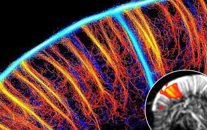
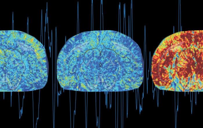
Dr. Pierre Pouget begins this webinar with an introduction to fUS brain imaging, including conventional doppler and ultrafast doppler imaging, permitting sensitive measurement of CBV and determination of functional hyperemia. A protocol for cerebral blood flow recording in the medial prefrontal cortex of Macaque monkeys during prosaccade and antisaccade tasks is illustrated, and the data collected shown. fUS enabled single trial detection of supplementary eye field activation with a high signal-to-noise ratio, and the change in CBV was measured.
“One pixel was even sometimes sufficient to detect whether the monkey was . . . waiting for the task or doing the task.”
Single cell electrophysiology was subsequently combined with fUS, and data collected during microelectrode implantation is shown, illustrating the benefit of using fUS to confirm placement for recording. Combining the different techniques enabled simultaneous collection of macroscopic fUS data, mesoscopic local field potential recordings, and microscopic single unit activation signals from the same brain region.
“These techniques allow us to not only access a large spatial region of interest but also be able to study this changing activity in a very short temporal and time resolution.”
Dr. Pouget concludes the first portion of the webinar with a summary of methods for observing the living brain, including ultrafast fUS, and the potential to combine these techniques with other existing methods.
In the second half of the webinar, Dr. Serge Picaud describes his interest in visual restoration strategies and the prevalence of age-related vision loss, including two of the major causes of blindness: macular degeneration, in which photoreceptors are damaged, and glaucoma, resulting in loss of retinal ganglion cells. Treatment options include activation of neurons with subretinal implants or direct activation of the visual cortex via cortical implants, but both approaches require a detailed understanding of the representation of visual information within the visual cortex.
“. . . we could use functional ultrasound imaging to assess these strategies for restoring vision.”
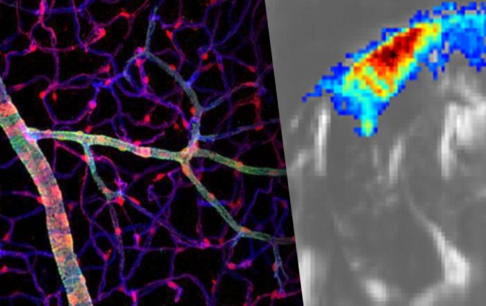
Initial experiments in rats presented with visual stimuli were conducted, during which activation of the visual system was recorded through a cranial window. Using this method, CBV in the superior colliculus, lateral geniculate nucleus, and the visual cortex (V1/V2) was measured, permitting 3D reconstruction of visual system activity and retinotopy of visual responses. Similar experiments were then performed in NHPs, demonstrating the quality of fUS images in comparison with MRI. The CBV response to signal presentation was measured, and clear responses were observed within only 10 trials. The data presented allowed for creation of retinotopic maps, including identification of ocular dominance columns in both superficial and deeper layers.
“. . . the first result of a patient who was able to, thanks to the activation of these retinal ganglion cells with optogenetic therapy, . . . find . . . some object on a table . . .”
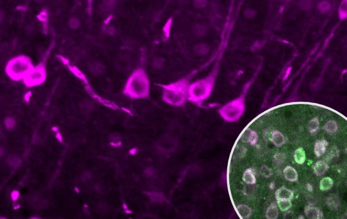
Examples of vision restoration clinical trials are then shown in which retinal prostheses were used in cases of age-related macular degeneration. This method was first developed in animal models, using isolated retinas and in vivo protocols, in which the response to infrared and visible light at different intensities was assessed by fUS and electrophysiological recording. The first clinical trials utilizing optogenetic therapy for vision restoration are also presented, together with new experiments exploring the use of optogenetic activation of neurons expressing ChrimsonR. Optogenetic activation was assessed simultaneously with fUS and electrophysiology, demonstrating a strong correlation between the two methods when measuring cortical activity.
Dr. Picaud concludes this webinar with a summary of these experiments and clinical trials, demonstrating the benefits of fUS to assess functional responses in the visual cortex.
Click to watch the webinar recording. To view the presentation full screen simply click the square icon located in the bottom-right corner of the video viewer.
Resources


Q&A
- What are the respective advantages of fUS, electrophysiology, and fMRI?
- What EEG frequency bands are most correlated with the fUS readout?
- Does the fUS signal correlate better with spikes or local field potentials?
- What would the fUS look like for finer stimuli and can you use this method for visual acuity cutoffs?
- What is the minimal stimulation duration?
- Will it be possible to do these types of recordings with the scalp intact?
- How are non-local blood flow changes (such as due to body movement) reflected in the fUS signal?
- How do you envision the clinical translation of these techniques?
- Do you have any data showing layer specific activation differences using fUS in the V1 or elsewhere?
- What are the future directions of this work?
To retrieve a PDF copy of the presentation, click on the link below the slide player. From this page, click on the “Download” link to retrieve the file.
Presenters
CNRS Principal Investigator
Brain and Spine Institute
Director
INSERM Vision Institute France
Production Partner
Iconeus