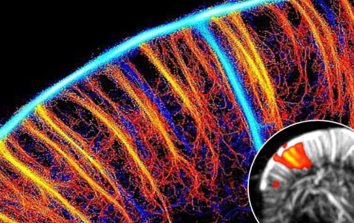
Dr. Marty Pagel introduces the CEST MRI technique, demonstrates a basic experiment, and explains why measuring protein, acid, and metabolite content can be so beneficial in applications of brain ischemia and cancer.
Key Topics Include:
- Introduction to CEST MRI for assessing disease parameters in brain cancer and ischemia
- How CEST compliments Bruker’s MRI/PET instruments
- Assessing tumor metabolism using CEST
- Bruker BioSpin’s new CEST method now available in ParaVision 360
- The upcoming CEST 2020 and CEST 2021 meetings, where more can be learned about this product and CEST MRI applications
Presenters

Professor
Departments of Cancer Systems Imaging and Imaging Physics
MD Anderson Cancer Center
Dr. Marty Pagel has focused on molecular imaging research during the last 20 years in industry and academia. He is a Professor in the Departments of Cancer Systems Imaging and Imaging Physics at the University of Texas MD Anderson Cancer Center. In addition, Dr. Pagel has held leadership positions in professional societies, funding agencies, and scientific journals that focus on molecular imaging. Dr. Pagel’s current research focuses on photoacoustic imaging, CEST MRI, DCE MRI, and PET/MRI, for studies with mouse models of cancer and for clinical trials with cancer patients.
Production Partner
Bruker Corporation
Bruker offers preclinical imaging solutions for a broad spectrum of application fields, such as cancer research, neuroimaging and cardiac disease.

Additional Content From Bruker Corporation
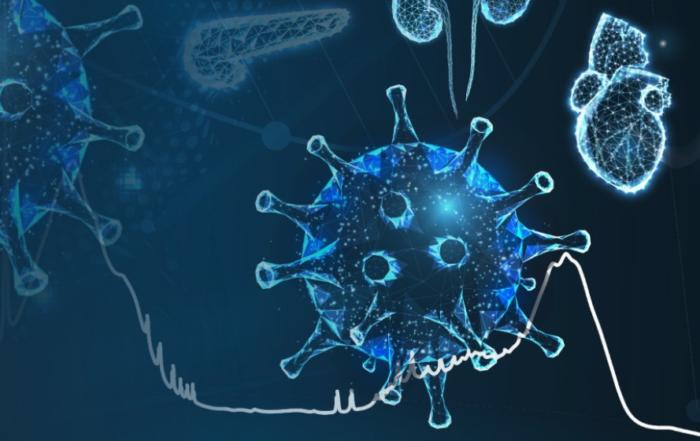
Unlocking Your Clinical Understanding of Post-Acute COVID Syndrome (PACS), Patient Recovery, and Risk of Subsequent Disease
Watch this webinar to learn about the metabolic changes during COVID-19 disease, how these can be accurately tracked and how NMR can improve the clinical understanding of Long-COVID.
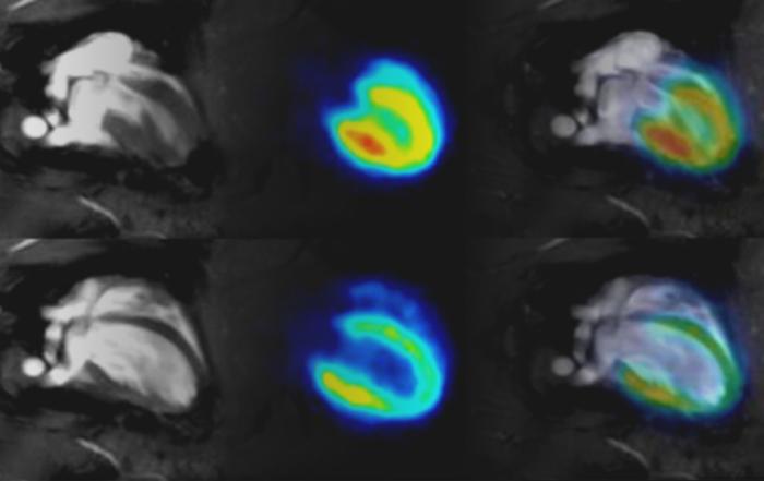
Cardiac PET/MR Imaging in Small Rodents
Experts provide a comprehensive overview of approaches to address cardiac and respiratory gating in PET and MRI in rodent models.
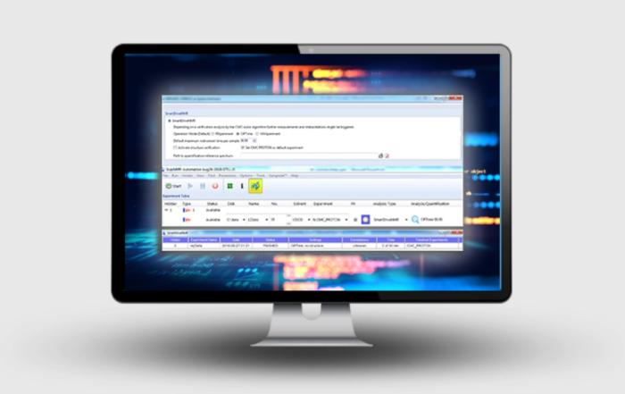
SmartDriveNMR – The Next Generation of Automation
Bruker experts provide an overview of SmartDrive with a particular focus on the exciting new features. They offer practical examples and demonstrate the newest features and functions live!
Related Content

Photoacoustic Imaging in Angiotensin II Induced Cardiac Hypertrophy Mice
Hear Dr. Emily Lupton on her lab's investigation whether photoacoustic imaging (PAI) could provide novel imaging in the heart after angiotensin-II induced hypertrophy with and without treatment with losartan.
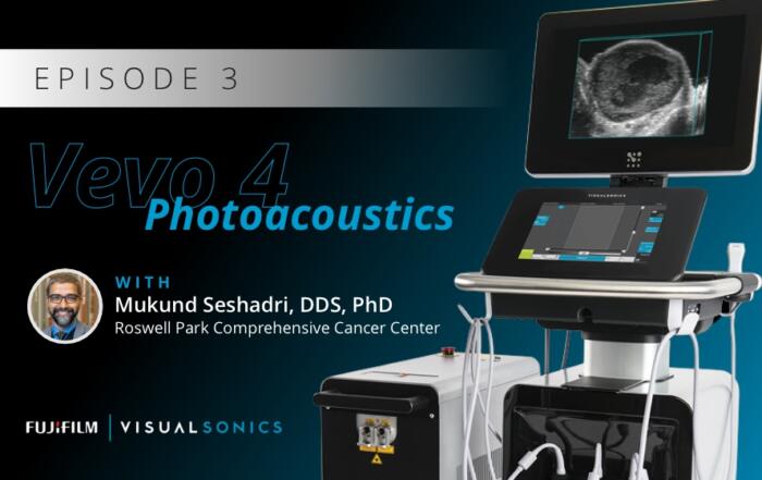
The Translational Utility of Ultrasound and Photoacoustics in Head and Neck Oncology
Dr. Mukund Seshadri presents his interdisciplinary research on developing and evaluating ultrasound and photoacoustic imaging for oral oncology applications, with a focus on the prognostic utility of these methods in assessing treatment responses in head and neck cancer patients.
Functional Ultrasound Neuroimaging: Principles, Applications, & Perspectives
Dr. Mickael Tanter presents his work on functional ultrasound neuroimaging, including an overview and translation to clinical applications.