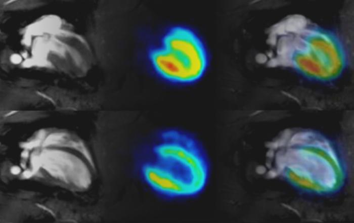
An infectious lung disease expert shares compelling evidence for the use of micro-CT to repeatedly evaluate disease progression and therapy efficacy in live animals.
Lung diseases such as fibrosis, viral infection and cancer are life-threatening conditions for which our knowledge on etiology and/or effective treatment options still contains important gaps. To unravel disease processes and to test novel therapeutic approaches, investigators typically rely on end-stage procedures (such as serum analysis, cyto-/chemokine profiles and selective tissue histology from animal models). These techniques are useful but provide only a snapshot of disease processes that are essentially dynamic in time and space. Technology allowing evaluation of live animals repeatedly is indispensable to gain better insight into the dynamics of lung disease progression and treatment. Computed tomography (CT) is a clinical standard and can have enormous benefits in a research context too. Yet, its implementation in basic and preclinical research laboratories lags behind. Nevertheless, the evidence for the benefits of micro-CT as an indispensable and safe tool to repeatedly evaluate lung disease progression and therapy efficacy in live animals is nothing but convincing.
Presenters

Assistant Professor
Imaging and Pathology Department and MoSAIC Imaging Core Facility
Ku Leuven
Production Partner
Bruker Corporation