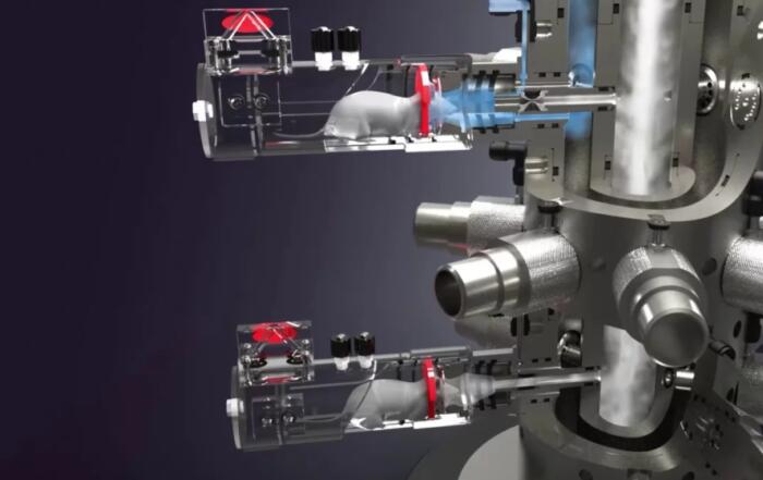
Dr. Redente discusses the advantages of microCT and provides a broad overview of pulmonary disease imaging using a highly innovate technique to analyze living animals, as well as fixed tissue.
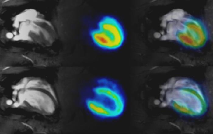
The ability to quantitate lung disease and monitor disease development and resolution in real time is an advantage to using small animal microCT. In addition, 3D analysis provides data about the whole lung instead of the snap-shots typically associated with conventional analysis techniques, including histology. microCT provides the ability to assess a large diversity of lung diseases and the outcomes correlated to data generated from human patients.
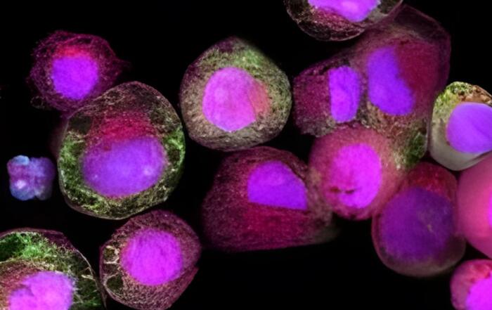
In this Bruker webinar, Dr. Elizabeth Redente, Associate Professor at National Jewish Health, will give insights from literature and her group on the ability of microCT imaging to quantitate, visualize and follow the development of a myriad of lung diseases including inflammation associated with infection, fibrosis, emphysema and tumor development. The use of 3-dimensional analysis to quantify lung disease is advantageous over traditional histology techniques that capture a single cross-sectional analysis or macro-dissection of the lung to isolate tumors, which reduces spatial understanding. In addition, the use of contrast agents allows for easy visualization and quantitation of organ vasculature.
Key Topics Include:
- MicroCT of the lung – Current methods and limitations
- Examples of lung disease
- Quantification and visualization for measuring lung volume, Hounsfield units, and tumour counting
- Benefits to using MicroCT
Presenters

Associate Professor of Paediatrics
National Jewish Health
Production Partner
Bruker Corporation