In this webinar, Dr. Anna Cavaccini and Dr. James Dooley share their insights into the development of rodent sensorimotor neuronal circuits during early postnatal life, including the cortical and subcortical mechanisms involved in the development of sensorimotor circuitry during wakefulness in mice and REM sleep in rats.
Highlights
- An introduction to the role of the striatum in movement during early development in mice
- Changes in locomotor activity and simultaneous measurement of striatal firing rates
- An introduction to forward models and their role in movement pathways
- The use of myoclonic twitches to study movement models in rats
Webinar Summary
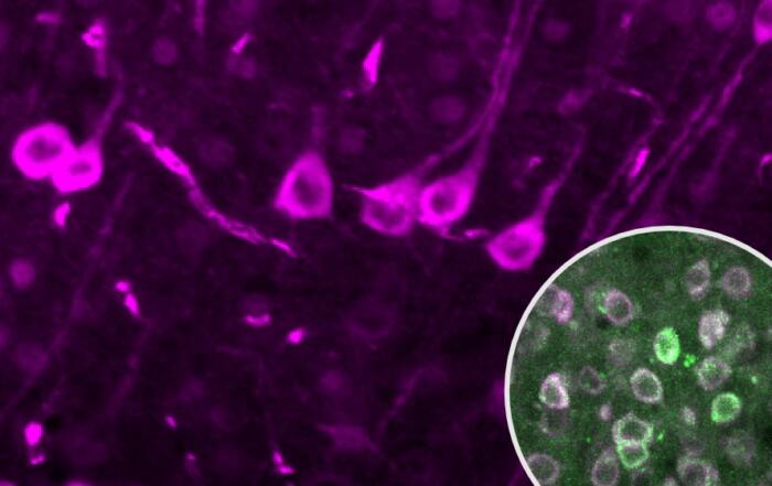
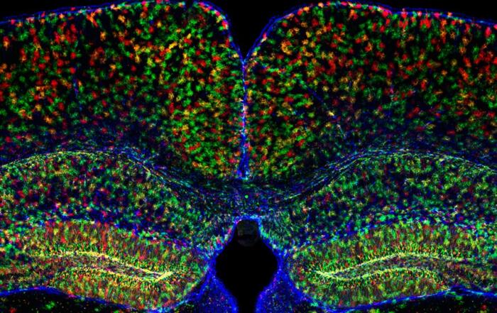
Dr. Cavaccini begins this webinar with an introduction to basal ganglia circuits and movement; although these pathways have been well-characterized in adult mice, less is known about their development during early postnatal life. Some differences in striatal circuitry have been reported in early mouse development, including increased excitatory synapse density, synaptogenesis, and morphological changes associated with striatal neurons; the aims of the experiments described in this webinar were to further investigate striatal output during development and evaluate the correlation between this output and locomotion.
“. . . striatal firing rate is decreased compared to young adult mice and this might suggest a different engagement of the striatum from the input . . .”
The challenges inherent in performing brain activity studies during locomotion in mouse pups are highlighted and how they may be overcome using the Neurotar Mobile HomeCage. In brief, the Mobile HomeCage was used to assess locomotion in freely-moving mouse pups in conjunction with silicon probe recording at different stages of development. Average movement speed increased over developmental stages together with travel distance and exploratory behavior, accompanied by an increase in striatal firing rate that was correlated with locomotor activity.
Dr. Cavaccini concludes the first portion of the webinar with a summary of the findings and describes a number of future experiments intended to further elucidate the relationship between striatal activity and locomotion during mouse development.
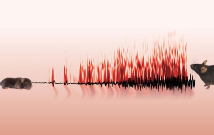
In the second portion of the webinar, Dr. Dooley begins with a number of videos demonstrating the complexity of sensory driven motor tasks to emphasize the question of how the brain addresses sensory and motor delays. One of the ways that the brain integrates this information is via a forward model that predicts sensory feedback from a given motor command with a time shift and inhibits sensory information due to the movement. Internal models of this type typically require large numbers of repetitions over prolonged periods, making study during development intrinsically difficult; myoclonic twitches are proposed as a means of overcoming this challenge.
“The cerebellum seems to be creating both a forward model and canceling expected sensory feedback.”
The experimental protocols are described, and included continuous data collection for several hours during sleep-wake cycles in rat pups using the Neurotar Mobile HomeCage with simultaneous electrophysiological recording from the ventrolateral and the ventral posterior nuclei of the thalamus. The data confirmed that myoclonic twitches are produced spontaneously in large numbers, are discrete and highly stereotyped, drive precise neural activity, and are represented by an internal model of movement. Dr. Dooley concludes this webinar with a summary of the experiments, and an outline of possible future studies to determine whether myoclonic twitches are necessary to establish and modify the underlying models of movement.
Click to watch the webinar recording. To view the presentation full screen simply click the square icon located in the bottom-right corner of the video viewer.
Resources

Q&A
- Does the specific engagement of striatal subpopulations during movement change over the course of development?
- Do twitches continue to calibrate the system?
- What thalamic nuclei are involved in motor control?
- Did you see any evidence of differences between the ventrolateral and the ventral posterior nuclei during waking movements?
- Have you stimulated cortical sensory areas to investigate striatal responses during development?
- How would the development of internal models apply to vision?
To retrieve a PDF copy of the presentation, click on the link below the slide player. From this page, click on the “Download” link to retrieve the file.
Presenters

Postdoc
Brain Research Institute
University of Zurich

Assistant Research Scientist
Psychological and Brain Sciences
University of Iowa
Production Partner
Neurotar