Dr. Yasaman Soudagar and Dr. Roshni Christo demonstrate how to simultaneously image calcium in up to 4 brain areas of freely behaving mice to map out neuronal circuits and identify changes that take place under different disease conditions and during different behavioral tasks.
Highlights
- A discussion of multi-region brain recording and imaging methods
- An introduction to the Neurescence Multiscope and experimental workflow
- A demonstration of imaging data collection and software
- Practical application of the Quartet system for simultaneous calcium imaging and behavioral testing in several experimental paradigms
- An overview of the Chromatone system for multiple wavelength illumination
Webinar Summary
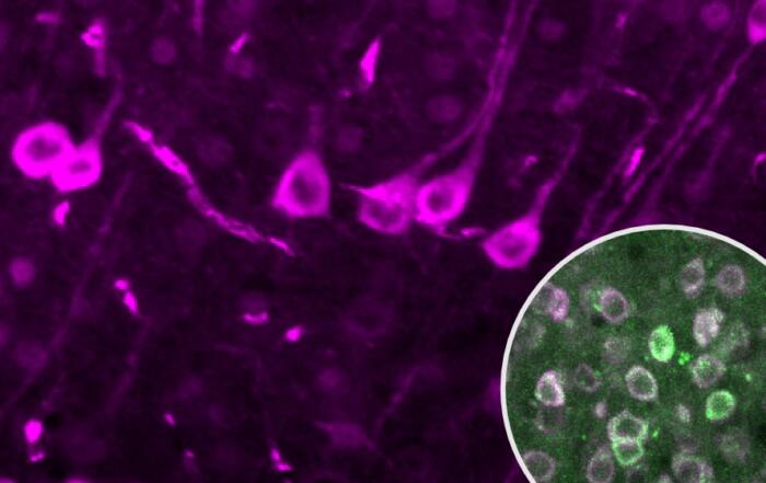
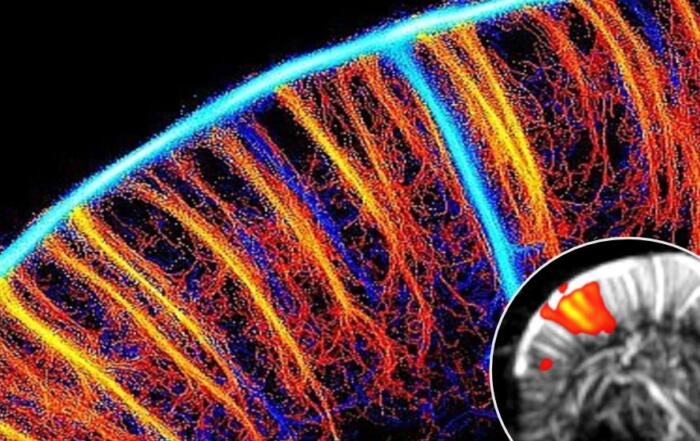
Dr. Soudagar begins this webinar by highlighting widespread interest in multi-region brain imaging across a diverse range of research programs. A number of fluorescent proteins have been developed for these applications, including GCaMP, a marker used to measure in vivo calcium signaling and as an indicator of neuronal activity. Currently available recording (electrophysiology and photometry) and imaging (2-photon and epi-fluorescence) methods are then summarized, along with the benefits, limitations, and costs associated with each of these approaches. Some of the key features of epi-fluorescence are that it allows for imaging in freely behaving subjects with access to deep brain areas, permits study of neuronal circuits, and does so at a significantly lower cost than other imaging modalities.
“There is really a large range of applications and a large range of questions that can be answered if one can simultaneously study multiple brain areas.”
The Neurescence Multiscope is a newly developed epi-fluorescence technique for multi-region brain imaging in freely behaving subjects and has been used in a number of different animal models. The Quartet system includes a small and lightweight fluorescence microscope, data collection software, and flexible fiber bundles with lens connectors for attachment to subjects. A workflow for possible experiments is described, beginning with transgenic models or viral injection for selective marker expression, lens implantation (using the Piccolo surgical accessory), and ultimately recording and behavioral testing. A behavioral testing setup is also shown as well as examples of imaging data collection, demonstrating the focus-locking feature of the system and the capabilities of the software.
“You can . . . image multiple days, even weeks, without having to refocus the system.”
In the second portion of the webinar, Dr. Christo discusses the specific application of the Neurescence Quartet system, in conjunction with behavioral paradigms, to study neuronal circuits in experimental models. In the first example, calcium imaging and task performance were assessed simultaneously using an elevated T-maze apparatus. The second example demonstrates bilateral hippocampus calcium imaging during an open field odor memory task, highlighting the ability of the animal to move freely during recording; this freedom of movement also enables use of the system during swimming tasks. Additional applications presented include social interaction studies with multiple animals in a single open field environment, recording from up to four individually-housed animals with a single system, and simultaneous electrophysiology data collection. The potential behavioral impact of multiple implants on a single mouse is also addressed, but due to the small size of the implants, no differences were observed.
“The optogenetics protocols are completely customizable.”
In the final portion of the webinar, Dr. Christo concludes by introducing the Chromatone, which allows for the use of three different illumination wavelengths per probe with customizable timing (continuous or pulse), as illustrated in a series of brief videos.
Click to watch the webinar recording. To view the presentation full screen, click the square icon located in the bottom-right corner of the media player.
Resources

Q&A
- Does the user need to manually define the ROIs for each cell?
- Can the Quartet system also be used in rats?
- How much does the implant weigh, and does it affect animal behavior?
- Can multiple implants be used in different brain regions in the same animal?
- How deep can the system reach?
- How long can the system be used in the same animal following implantation?
- Are there any publications demonstrating the use of this system in the spinal cord?
- Are anesthetics necessary, and if so, how do they affect the results?
- Are there any issues with scarring after surgery obscuring the focal plane or changing the field of view?
- Are there different probe sizes or plans to introduce different probes for different applications?
- How much does the system cost?
- Can this system be used in areas other than the brain, and if so, how would they be fixed in place?
To retrieve a PDF copy of the presentation, click on the link below the slide player. From this page, click on the “Download” link to retrieve the file.
Presenters

Application Scientist
Neuroscience
Neurescence Inc

CEO
Neurescence Inc.
Production Partner
Neurescence Inc.