Join Mark Nielsen for a discussion on the evolutionary and developmental patterns that clarify the structural organization of the peripheral nervous system. This is the second webinar in this 4-part series on how science education has evolved in the face of new challenges.
The peripheral nervous system, from the cranial nerves to the spinal nerves and all their autonomic branches, is a daunting network of wires that communicate input and output between all parts of the body and the central nervous system.
Key Topics Include:
- Why do all spinal nerves have the same basic structure?
- Why does spinal nerve structure differ from cranial nerve structure?
- Why are there dorsal root ganglia and not ventral root ganglia?
- Why are there autonomic ganglia and two efferent neurons in the autonomic pathways?
- Why is the parasympathetic output craniosacral in origin and the sympathetic output thoracolumbar in origin?
- Why are there white communicating rami at only some spinal nerve levels and gray communicating rami at all spinal nerve levels?
- Which nerves retain a more primitive structure, cranial nerves or spinal nerves and why?
Who Should Attend?
This presentation is intended for professors who want to have a more thorough understanding of peripheral nervous anatomy so they can more effectively teach students about this network of nerves and help their students become more effective learners.
Click to watch the webinar recording. To view the presentation full screen simply click the square icon located in the bottom-right corner of the video viewer.
Resources

Presenters

Professor, John Legler Endowed Lecturer of Human Anatomy
Biology
University of Utah
Mark Nielsen is a Professor in the Department of Biology at the University of Utah. For the
past thirty-five years he has taught anatomy, neuroanatomy, embryology, human dissection,
comparative anatomy, and an anatomy teaching course to over 28,000 students.
His graduate training is in comparative anatomy, and his anatomy expertise has a strong
basis in dissection. He has prepared and participated in hundreds of dissections of both humans
and other vertebrate animals. All his courses incorporate a cadaver-based component to
the training with an outstanding exposure to cadaver anatomy. He is a member of the American
Association for Anatomy (AAA), the Human Anatomy and Physiology Society (HAPS) of which
he is the past president, and the American Association of Clinical Anatomists (AACA).
Production Partner
ADInstruments
Established in 1988, ADInstruments develops high performance digital data acquisition and analysis solutions for biomedical research and life science education.

Additional Content From ADInstruments

Getting to the Heart of Cardiovascular Research: From Yesterday to Today and Looking Towards Tomorrow
Dr. Melanie White presents the various methods for assessing cardiac function in the context of pathology, spanning from in vitro to in vivo techniques, and how she integrates these with cutting-edge mass spectrometry in her research on cardiovascular disease pathogenesis.
Evaluation of Novel Therapies Using Spontaneous Seizure Models
In this webinar, Dr. Cameron Metcalf covers the technical requirements and benefits of spontaneous seizure models, addressing the challenges and optimization of 24/7 video-EEG data collection for epilepsy therapy development.
Measurement of Cardiac Function Using Pressure-Volume Loops in Swine
Dr. Pedro Ferreira gives a deep dive into his work using pressure-volume loops in large animal models of heart failure.
Additional Content From Human Anatomy & Physiology Society
Creating Community in Online STEM Classes
Wendy Riggs, MS discusses the difficulties of building a community in online classrooms and how to overcome these barriers using technology.
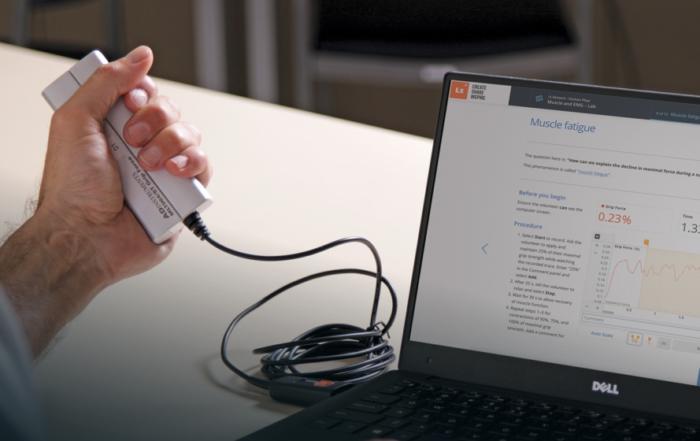
Examining the Anatomy and Physiology Lab Experience
Join Wendy Riggs for a deep dive into the virtual Anatomy and Physiology Labs that she runs at College of the Redwoods and how she uses technology to facilitate meaningful learning opportunities for her students.
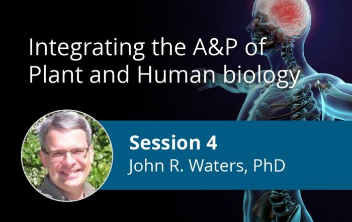
My Cousin, the Tree: Integrating the Anatomy & Physiology of Plant and Human Biology
Join Dr. John Waters as he discusses how to help students organize their understanding of biology around larger themes that are common across the life sciences. This is the fourth webinar in this 4-part series on how science education has evolved in the face of new challenges.
Related Content
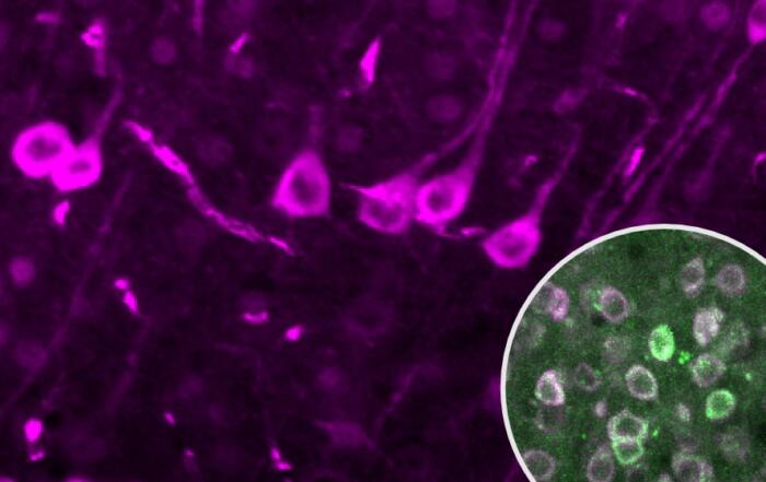
Spatiotemporal Dynamics of Calcium and Neurotransmitters in Awake Mouse Models of Epilepsy
In this webinar, Dr. Vincent Magloire and Dr. Ken Berglund present their work on the spatiotemporal dynamics of neurotransmitter and calcium imaging during seizure evolution in awake head-fixed mice and models of epilepsy.
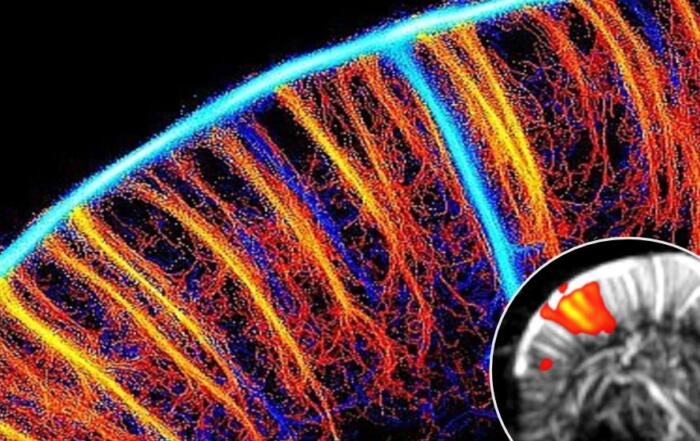
Functional Ultrasound Neuroimaging: Principles, Applications, & Perspectives
Dr. Mickael Tanter presents his work on functional ultrasound neuroimaging, including an overview and translation to clinical applications.

Updates in Chronic Traumatic Encephalopathy (CTE)
Dr. Ann McKee will describe the emergence of chronic traumatic encephalopathy (CTE) as a distinct disease over the past 20 years.