Q&A Report: Unattended FIB Serial Sectioning Tomography for 3D Characterization Using Auto Slice and View 4
The answers to these questions have been provided by:
Brandon Van Leer, MS
Applications & Business Development,
Materials & Structural Analysis,
Thermo Fisher Scientific
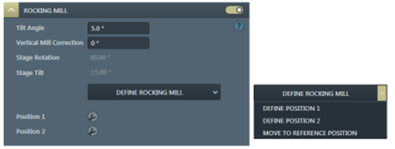
1. How can one improve 3D Characterization Quality when using a Plasma FIB on materials that curtain very easily?
Auto Slice and View 4 comes standard with technology that allows the user to mitigate curtaining during focused ion beam serial sectioning tomography (FIB-SST). Thermo Fisher Scientific has embedded a rocking polish application into the software that mitigates and even eliminates curtain formation during the sectioning routine. This application is customizable by the user so that rocking polish is optimized for the best results. (Reference Graphic Attached).
2. How does Auto Slice & View 4 maintain the best imaging results during acquisition?
Auto Slice and View 4 offers the user advanced beam control settings to ensure the best image for each slice. Automated functions include: Auto-focus, auto-stigmation, auto-lens align and auto-source tilt. Each of this automated functions can be run for each slice or customer to be run every nth slice. The choice is up to the user. Additional parameters for each of these automated functions are available during imaging set-up and can be changed on-the-fly during the acquisition.
3. Is it possible to control the microscope and Auto Slice and View remotely?
Thermo Fisher Scientific has enabled remote control of our scanning electron and DualBeam FIB-SEM microscopes for more than a decade. Because the microscopes are fully digital, it is possible to control the system remotely with just a mouse and keyboard. Often times, our own applications personnel take advantage of this capability to work remotely on demonstration systems or even as a training opportunity with our customers.
4. Is it possible to change the stage position of the sample during an acquisition to take advantage of improved detection and imaging performance with the SEM at an optimized sample position?
Yes, Auto Slice and View allows the user to optimize the FIB location for serial sectioning and the SEM imaging location for best imaging results (if desired). The application saves the position for SEM and FIB separately. Using image matching with a fiducial ensures the sample is in the correct location each time. Taking advantage of this capability allows the user to even use retractable, in-chamber detectors like a dedicated solid-state diode backscatter electron (BSE) detector when desired.
5. With plasma FIBS, what is the smallest achievable slice thickness in a 3D tomography?
Because plasma FIBs have different source technology than a traditional liquid metal-ion source (one of the benefits that allows them to achieve beam currents 20X larger than the former mentioned sources), their inherent resolution is also about 5X worse. However, for FIB-SST and even S/TEM sample preparation, there is more than enough resolution to achieve the desired results. PFIB specialists at Thermo Fisher have demonstrated slice thickness as small as 10 nm using 30 kV accelerating energy. For cross-sectional areas greater than 100 X 100 µm^2, slice thickness as low as 50 nm is achievable.
6. Can one acquire both secondary and backscattered electron images from the same cross-section face? What about EDS?
Yes, Auto Slice and View allows the user to acquire multiple signals from the same cross-section face. In some cases, these signals can even be acquired simultaneously to save acquisition time. For example, if the user would like to acquire both a secondary electron and backscatter electron image, the application allows the user to acquire both of these signals simultaneously from two different detectors. It is even possible to cut with the FIB and then take a secondary electron image generated by the focused ion beam (instead of the SEM). If, after image acquisition, the user would also like to acquire analytical data, the user can also generate this data on the same cross-section face prior to the next serial sectioning cut with the FIB. In fact, the user can customize acquisition for different signals to happen on each slice for every nth slice.
7. What is the typical time it takes to acquire a 3D dataset in the microscope?
Typical acquisition times for a 3D acquisition run from a few hours to several days. It depends on how large of a volume a user has a desire to characterize by FIB-SST. Typical acquisition times for an electron image run with no analytical results span from 12 hours to approximately 48 hours. If analytics are desired, acquisition times can extended to 3 or 4 days depending on the volume of material.