Join Professor Annarosa Arcangeli as she discusses the use of multi-modal imaging to detect and monitor pancreatic and liver tumors.
In oncology, optical imaging provides only one piece of the puzzle, with new methods and approaches continually being developed to provide additional information. In the first episode of the Vevo 4 Oncology Web Series, Prof. Arcangeli discusses a new and powerful, high resolution multi-modal imaging solution that combines ultra high frequency ultrasound and photoacoustic imaging. Several applications of this system are presented, including tumor/polyp detection and sizing in 2D and 3D, detection and monitoring of tumor growth and angiogenesis, as well as others.
Key Topics Include:
- Tumor cell guided injection
- Tumor/polyp detection and sizing in 2D and 3D
- Detection and monitoring of tumor growth and angiogenesis
- Pancreatic tumor perfusion and volume quantification
- Contrast-enhanced imaging of biomarkers
- Coregistration of ultra-high-resolution ultrasound and photoacoustic imaging to detect tumor oxygen saturation
Presenters

Professor
Department of Experimental and Clinical Medicine
University of Firenze, Italy
For almost thirty years, Dr. Arcangeli has been involved in studies aimed at defining the biophysical aspects of intracellular signaling by controlling cell growth and differentiation of tumor cells. She contributed to unravel the action mechanism of widely used inducers of tumor cell differentiation, such as the "hybrid polar compounds" (HPC). She also focused on the role of potassium channels, in particular, the hERG1 channel, in the governance of the resting potential, cellular ionic homeostasis and cell signaling in normal and cancer cells.
Webinar Host
FUJIFILM VisualSonics Inc.
FUJIFILM VisualSonics designs and manufactures ultra high frequency in vivo imaging systems, for both research and clinical use. our ultrasound platform provides images at resolutions that far exceed any other system available on the market. Beyond ultrasound, we have also developed a unique photoacoustic technology to expand on the capabilities of our imaging solutions.

Additional Content From FUJIFILM VisualSonics Inc.

Photoacoustic Imaging in Angiotensin II Induced Cardiac Hypertrophy Mice
Hear Dr. Emily Lupton on her lab's investigation whether photoacoustic imaging (PAI) could provide novel imaging in the heart after angiotensin-II induced hypertrophy with and without treatment with losartan.
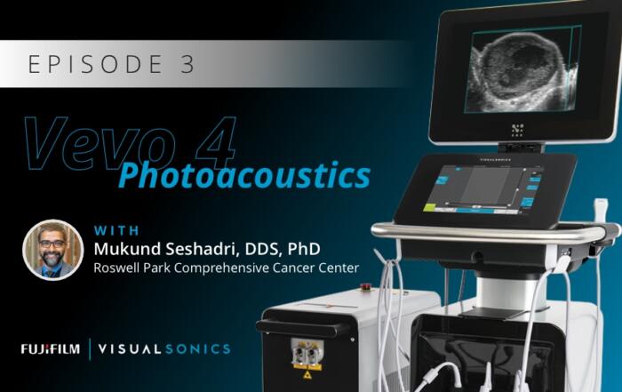
The Translational Utility of Ultrasound and Photoacoustics in Head and Neck Oncology
Dr. Mukund Seshadri presents his interdisciplinary research on developing and evaluating ultrasound and photoacoustic imaging for oral oncology applications, with a focus on the prognostic utility of these methods in assessing treatment responses in head and neck cancer patients.
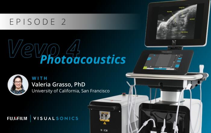
Superpixel Unmixing Algorithm from Micro to Macro Photoacoustic Imaging of Tissue Components
Dr. Valeria Grasso describes a novel unsupervised Machine-Learning method named Superpixel Photoacoustic Unmixing (SPAX) framework.
Related Content

Circulating Nucleosome Quantification (Nu.Q®) Technology for Monitoring Target Engagement and Adverse Events in Oncology Trials
Join us for a comprehensive webinar featuring experts in epigenetics and assay development, focusing on the scientific foundations and applications of Nucleosome Quantification (Nu.Q®) Technology in the realms of drug development and disease monitoring.
Assessing Antigen-Specific T-Cell Functionality with Dendritic Cell/CD8⁺ T Cell Co-culture
In this webinar, learn how to set up DC and CD8⁺ T cell co-culture experiments and assess T cell proliferation and killing activity.
A Ready-to-Analyze High-Plex Spatial Signature Development Workflow for Cancer Immunotherapy
In this webinar, Aditya Pratapa and Lorcan Sherry present a new workflow for analyzing multiplex immunoflurescence images.