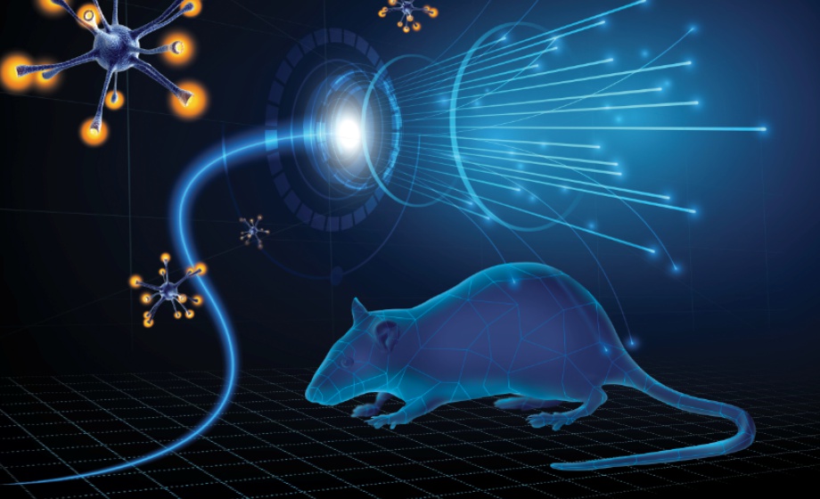
Q&A Report: Optogenetic Activation of Selective Cardiac Autonomic Neurons
What are the current limitations and challenges of cardiac optogenetics?
Although new and improved opsins are constantly being developed, the main limitation of optogenetics is the relatively reduced level of tissue penetration, notably in dense cardiac tissues as a result of increased light attenuation and scattering. If stimulated from the epicardial surface, this will specially limit the access to certain endocardial sites that could provide greater insight into cardiac conduction pathways.
One of the greatest challenges of cardiac optogenetics is how to translate it from animal studies to human clinical applications. When considering the different methods to induce a uniform opsin expression in human cardiac tissue, there is a special concern for cardiac toxicity and immunogenicity associated with opsin expression and viral capsids that could jeopardize optimal opsin delivery. This could bring additional concerns; particularly, a nonuniform expression could result in heterogeneous cell depolarization/hyperpolarization that could eventually lead to arrhythmias. Moreover, uniform photo-stimulation throughout the entire heart presents an additional technical challenge. In this case, given the vigorous contraction of the heart and the lack of easy access to the cardiac surface, novel custom-made biocompatible electronics will likely be required that fully adapt to the morphology of the heart while adjusting the overall energy for optogenetic stimulation.
Is it possible to use optogenetics to stimulate non-neuronal cardiac cells?
Even though optogenetics was initially implemented for neuroscience, the field has evolved exponentially to the point of reaching different cells involved in cardiac control and sensing. For instance, different studies have shown ChR expression in cardiomyocytes, embryonic stem cell-derived cardiomyocytes, and fibroblasts for cardiac pacing, and arrhythmia control and termination.
A couple of examples of such applications can be seen in the following articles:
Which opsins are most suitable for cardiac optogenetic control?
There are many different opsins with unique biophysical properties that are suited to specific applications. For optogenetic control of cardiac function, there are two main types: the excitatory or depolarizing opsins such as Channelrhodopsin (ChR) which are ion channels with relatively fast kinetics and capable of generating enough photocurrent to elicit an electrical response in excitable cells, and the inhibitory or hyperpolarizing opsins such as the chloride pump Halorhodopsin (HR) and the proton pump Archaerhodopsin (AR) with the potential of suppressing or limiting excitability in cardiac cells.
Several of these opsins have evolved into versions with improved performance. The larger photocurrent and greater conductivity mutation H134R in ChR2, the red light-shifted ReaChR that provides deeper tissue penetration, the enhanced calcium permeability CatCh, and the Arch-T with a relatively large hyperpolarizing current, are just some examples of the different capabilities of the many opsins available.
In the end, the type of opsin to use for cardiac optogenetic control will highly depend on the application and the specific requirements.
For more information, please check these review articles:
How close should the LED be to the neurons you want to stimulate? Should it be directly above them, or can it be 2 mm to the side?
This will depend on the autonomic branch you want to stimulate. The most important aspect is to be able to stimulate specific regions, i.e. when stimulating the parasympathetic branch is preferably to place the LED on the right atrium, facing the SA node, whereas for the sympathetic branch, we have a wider range because there are nerves that innervate the atria and ventricles. By considering this, LEDs (particularly microLEDs) that are directly placed on top of the area where the neurons are, will have a narrower beam and a much more focused illumination pattern compared to an LED that is located ≥2mm away from the target. Thus, for parasympathetic stimulation is preferred to have the LED as close as possible to the heart to avoid illuminating unintended regions, and for sympathetic stimulation it is possible to even use a spotlight LED that is farther away from the heart. However, regardless of the distance of the LED, it is important to maintain the minimum irradiance necessary to induce a response (in our case was about 0.68 mW/mm2). If the power that feeds the LED is constant, the irradiance will change with distance.
Why is red light better for cardiac cell stimulation?
Red light is not necessarily better for cardiac cell stimulation; it will depend on the type of application that is needed and the required wavelength. However, red light-shifted opsins do provide certain advantages with respect to other opsins that require, for instance, blue light for stimulation: because red light has less scattering and absorption in tissue, it has a deeper penetration capability into cardiac tissues, requires less irradiance for excitation, and could potentially exhibit larger photocurrent.
The Red-activatable Channelrhodopsin (ReaChR) is an excellent example of the capabilities of a red light-shifted opsin. For more information on its properties and potential applications for cardiac control, please check the following articles:
When you verify gene expression with confocal microscopy, do you take thin heart tissue slices for immunofluorescence? Do you stain cardiomyocytes for innervation location of neurons?
Certainly, for confocal microscopy we take thin slices (~100 µm) from the base of the heart for sympathetic neurons, and from the right atrium for parasympathetic neurons. To localize the specific set of neurons, heart tissues are stained with primary and secondary antibodies: for sympathetic neurons, the slices are co-stained with rabbit anti-TH primary antibody, and then with goat anti-rabbit Cy5 and donkey anti-chicken Dylight 405 secondary antibodies; for parasympathetic neurons, the sample is placed in goat anti-ChAT primary antibody, followed by Alexa Fluor 647 conjugated donkey anti-goat secondary antibody.
For details of the entire process, please check these articles: