VisualSonics presents the first episode of their Vevo 4 Photoacoustics Web Series.
Combined Near Infrared Photoacoustic Imaging and Ultrasound Detects Vulnerable Atherosclerotic Plaque with Martin Schneider, MD, PhD from Stanford University.
Key Topics Include:
- Visualization of atherosclerotic plaque components with high-frequency ultrasound and photoacoustics
- Identification of photoacoustic biomarkers for atherosclerosis
- Distinguishing stable from vulnerable plaques
- Clinical significance of novel photoacoustic-detection methods
Who Should Attend?
Open to those studying vascular diseases or those interested in learning about new methods of non-invasive molecular imaging (all educational/experience levels are invited).
Presenters
Postdoctoral Fellow
Radiology
Stanford University School of Medicine
Martin Schneider joined the Gambhir Lab at Stanford in 2019 where he worked on optical phantoms and clinical photoacoustic imaging trials. He received the Cardiovascular Award from FUJIFILM VisualSonics.
Webinar Host
FUJIFILM VisualSonics Inc.
FUJIFILM VisualSonics designs and manufactures ultra high frequency in vivo imaging systems, for both research and clinical use. our ultrasound platform provides images at resolutions that far exceed any other system available on the market. Beyond ultrasound, we have also developed a unique photoacoustic technology to expand on the capabilities of our imaging solutions.

Additional Content From FUJIFILM VisualSonics Inc.

Photoacoustic Imaging in Angiotensin II Induced Cardiac Hypertrophy Mice
Hear Dr. Emily Lupton on her lab's investigation whether photoacoustic imaging (PAI) could provide novel imaging in the heart after angiotensin-II induced hypertrophy with and without treatment with losartan.
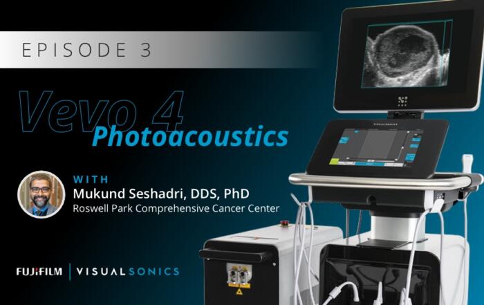
The Translational Utility of Ultrasound and Photoacoustics in Head and Neck Oncology
Dr. Mukund Seshadri presents his interdisciplinary research on developing and evaluating ultrasound and photoacoustic imaging for oral oncology applications, with a focus on the prognostic utility of these methods in assessing treatment responses in head and neck cancer patients.
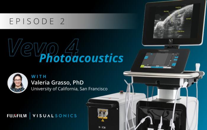
Superpixel Unmixing Algorithm from Micro to Macro Photoacoustic Imaging of Tissue Components
Dr. Valeria Grasso describes a novel unsupervised Machine-Learning method named Superpixel Photoacoustic Unmixing (SPAX) framework.
Related Content
Functional Ultrasound Neuroimaging: Principles, Applications, & Perspectives
Dr. Mickael Tanter presents his work on functional ultrasound neuroimaging, including an overview and translation to clinical applications.
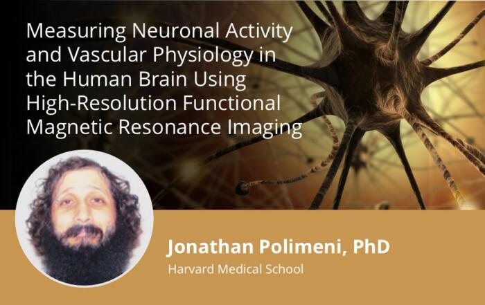
Measuring Neuronal Activity and Vascular Physiology in the Human Brain Using High-Resolution Functional Magnetic Resonance Imaging
Join Jonathan Polimeni, PhD for a deep dive into the physics and physiology of fMRI.
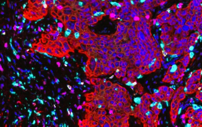
A Ready-to-Analyze High-Plex Spatial Signature Development Workflow for Cancer Immunotherapy
In this webinar, Aditya Pratapa and Lorcan Sherry present a new workflow for analyzing multiplex immunoflurescence images.