Join Jonathan Polimeni, PhD for a deep dive into the physics and physiology of fMRI and an understanding of its use for the study of neurological disorders.
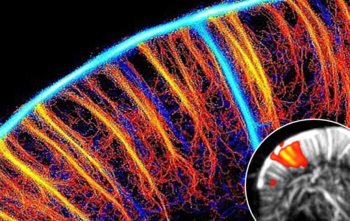
Functional MRI (fMRI) is the most widely-used method for measuring neuronal activity noninvasively across the entire human brain. All fMRI techniques in use today measure brain function indirectly—by tracking the changes in blood flow, volume, and oxygenation that accompany neuronal activity, and this has often been viewed as the fundamental limitation of the technique. However, recent evidence from microscopy studies has shown that the smallest blood vessels of the brain respond far more precisely, in space and in time, to neuronal activity than previously believed. This insight suggests that the “biological resolution” of fMRI is intrinsically high, and, with sufficiently high imaging resolution, it should fundamentally be possible to extract meaningful neuronally-specific information from fMRI—provided that we understand how vascular anatomy and physiology shape the hemodynamics that generate the fMRI signals.
This lecture describes ongoing efforts to improve the neuronal specificity of fMRI and pose the question: How far can we go with fMRI? A key challenge is that the vascular architecture of the brain reflects its structure and function across spatial scales. Modern fMRI is also used to measure cerebrovascular physiology, which is impaired in many neurological disorders, and so a deeper understanding of the fMRI signals will improve our understanding of the brain in health and disease.
Key Topics Include:
- How functional Magnetic Resonance Imaging (fMRI) measures brain function by tracking hemodynamic changes caused by active vascular responses to neuronal activity
- What is our current understanding of the spatial and temporal resolution of hemodynamics-based functional imaging methods like fMRI
- How vascular anatomy and physiology shape or influence the fMRI signals, and how this can be taken into account when analyzing and interpreting fMRI data
- How explicit models of realistic vascular anatomy and dynamics can help infer underlying neuronal activity using fMRI
- How fMRI can be used to measure vascular dysfunction in neurological disease
Presenters

Associate Investigator/Associate Professor of Radiology
Athinoula A. Martinos Center for Biomedical Imaging
Massachusetts General Hospital/Harvard Medical School