Q&A Report: Studying Epilepsy in Awake Head-Fixed Mice Using Microscopy, Electrophysiology, and Optogenetics
Can you image calcium transients in microglia while the animals are seizing?
A. Umpierre: Overall, yes, you can image microglial calcium transients during seizure. Seizures often result in X/Y axis oscillations, which can be corrected through motion-correction software. On rarer occasions, some Z-axis oscillation can occur, which cannot be corrected, requiring frame removal.
What is the propagation velocity and duration of the calcium spreading waves?
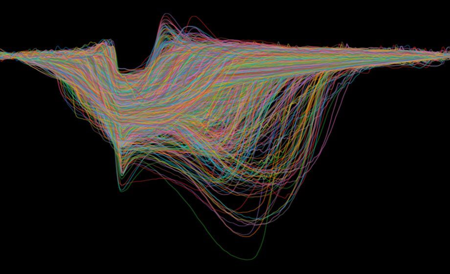
A. Umpierre: On average, microglial calcium waves spread between 1-3 microns/second. They can spread throughout the area of imaging, representing at least 150 microns of radial spread. The duration of the spreading calcium wave shows many microglial cells involved sustain calcium elevations for 3-4 minutes, with some sustaining calcium activity up to 8-9 minutes.
Is it possible that microglial Ca2+ activity in post-status period is due to neuronal activity?
A. Umpierre: Based upon our methodology, this is not possible to know definitively. We did not record simultaneous local EEG with our two-photon imaging, meaning we could not know in real-time if calcium events co-occurred with neuronal activity increases. The microglial calcium literature in vivo suggests that inflammation is also a strong driver of microglial calcium activity and could certainly contribute to the longer-term calcium elevations observed following kainate status epilepticus. In general, studies of cortical EEG in this model suggest that interictal spiking is pretty common in hippocampus and cortex during the time course imaged post-status. Broadly, then, signatures of aberrant neuronal network activity should have been present during the imaging period, but were not studied directly.
How do you deal with convulsive seizures? Do you correlate behavioral seizures and calcium events?
A. Umpierre: Convulsive seizures were really only considered during status epilepticus. They are predicted to be rare-to-absent in the 14 days post status epilepticus studied longitudinally in this model. We have recently adopted new capabilities to correlate these events: (1) the motion-tracking platform can give information about immobility during seizure; (2) an infrared camera inside the two-photon cage can give the most direct information about behavioral seizures, including the ability to perform Racine scoring; (3) implant of a cortical EEG system in current pilot studies allows us to record the electrographic signatures of a seizure in real-time along with the two-photon calcium imaging. In the published study, we did not have the capability to correlate behavioral seizures and calcium events definitively (no camera or EEG). Instead, we performed visual monitoring for seizures and then began imaging.
Is there always a general delay of 10 minutes to detect microglia activity?
A. Umpierre: Yes. There seems to be a 6-15 minute delay in all experiments in which we could accurately establish a delay latency (beginning from the moment the mouse was induced by isoflurane, or the time after peak neuronal activity changes following CNO). This appears to be a delay associated with microglial responses to neuronal activity changes. Damage responses, on the other hand, can occur as real-time calcium activity following injury. For example, Eichhoff et al. (2011) showed that destruction of a single neuron could elicit near-immediate calcium responses in local microglia.
How can we use head-fixed preparations to study spontaneous seizures in chronic models of TLE?
R. Wykes: Yes, but there are a couple of issues to consider. Spontaneous seizures, by definition are random. We normally restrict head-fixation experiments to ~2hrs. Although it is possible to insert a probe into the hippocampus and detect spontaneous seizures in mouse models of TLE, the number of successful recordings sessions where a seizure is detected is low. Additionally, it is possible to calcium image activity from the hippocampus, but this involves more invasive surgical preparation prior to experimentation.
Could a seizure lead to implant detachment?
R. Wykes: We have not observed this. In the chemoconvulsant models we use there are frequent brief (~10s) seizures that are correlated with increased running speed and eye pupil dilation. In a ~90 min recording session we usually detect 2-5 longer-lasting (>60s) seizures that generalize. These are associated with more pronounced behavioral manifestations. However the head bars remain attached.
Have you thought about designing a wireless system to eliminate limitations of head fixation?
R. Wykes: Yes, for chronic electrographic recordings we are utilizing a wireless head stage made by multichannel systems to couple to the graphene probes via a ZIF connected to a PCB. We have not considered a wireless system for calcium imaging so far.
How do you distinguish an invading ictal wavefront from a CSD? Is it just speed of propagation? Timing relative to LFP near the PTX injection site?
R. Wykes: Speed is the most obvious distinction. Seizures propagate ~0.5mm /s whereas CSDs propagate at 2-5mm/min. Additional seizure activity oscillates and can follow non-contiguous pathways, whereas CSDs appear, at least in this model to spread radially with no apparent respect to the underlying functional connectivity of the cortex.
Do you have a quantitative manner to determine if a mouse is well habituated?
Neurotar team: At Neurotar, we rely on behavioral o bservation, focusing primarily on the
tail position (up or down) and the locomotive patterns (jerky or relaxed). For measurements
of stress hormones in the Mobile HomeCage, see a recent publication from our user lab K.
Juczewski et al. (Scientific Reports, 2020). Our training protocol includes two days of
acclimation, two days of habituation, and two to three days of training. The training consists
of 2 3 brief daily sessions (5 10 min). This training duration is sufficient for most mice, but
there is variability between individual animals. Some mice require 5 7 days of training.
What are the minimum requirements for this equipment?
Neurotar team:
(1) Airflow: The Mobile HomeCage (standard size) requires an airflow of 120 L/min at 20 kPa, and the Mobile HomeCage Large – 150 L/min at 20 kPa. If your local air source does not satisfy these requirements, Neurotar’s team can recommend commercial air pumps.
(2) Compatibility with two-photon set-ups: The standard-size Mobile HomeCage fits into the majority of commercial two-photon microscopes. The Mobile HomeCage Large is compatible with most of the in vivo two-photon imaging systems. For assessing the compatibility with your imaging set-up, please refer to https://www.neurotar.com/taking-measurements-for-compatibility/ or contact us at mhc-support@neurotar.com
3) Stage: The XY translation stage must be robust. The Mobile HomeCage’s weight ranges between 6-13 kg, depending on the model.
(4) PC hardware and software requirements (for locomotion tracking):
OS: Windows 7 x64 or Windows 10 x64. 32-bit versions are not supported.
CPU (desktop): any processor with four real cores (e.g., AMD Ryzen 3 1300 or Intel Core i5 6400 and up).
CPU (mobile/laptop): most of the modern AMD Ryzen and Intel Core processors with four real cores (caution: large variability between processors depending on laptop model)
RAM: at least 4 GB, preferably 6 GB.
Storage: software and drivers require approx. 2 GB of disk space. Recorded tracks require up 100 MB per hour.
Ports: one type-A USB 3.0 port.