Dr. Matt Silva showcases the power of cryo-fluorescence tomography (CFT) in advancing immunotherapy research.
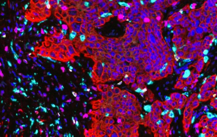
Cell-based immunotherapy is an emerging development in personalized cancer treatment where one application includes utilizing engineered immune cells that target cancer cells and engage the immune system to eradicate the disease. During preclinical development of these immunotherapies, it is essential to visualize cell distribution to both target and non-target tissues as well as gain critical information on immune engagement.
In this webinar, Dr. Matt Silva, CEO of Emit Imaging shows how whole-body cryo-fluorescence tomography (CFT) imaging will revolutionize cell and immunotherapy research. CFT enables multiplexing of cell tracking and localization with target and non-target tissue with immune activation via constitutive or imaging probe fluorescence. He covers the fundamentals of CFT imaging via the Emit XerraTM system and reviews examples of recent work on the platform.
Key Topics Include:
- Introduction to Cryo-Fluorescence Tomography (CFT) for immunotherapy research
- Overview of capabilities of the Emit Xerra system
- Real-world examples and applications of CFT for cancer research
Resources


Presenters

CEO
EMIT Imaging