Mapping Heterogeneous Interfaces Using Single-Entity Electrochemical Microspectroscopy
Dr. Venky Prabhakaran describes the development of scanning electrochemical microspectroscopy and its application to characterize heterogeneous electrified interfaces.
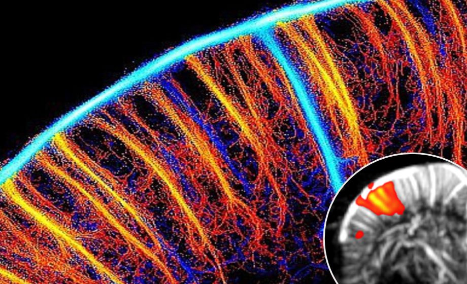
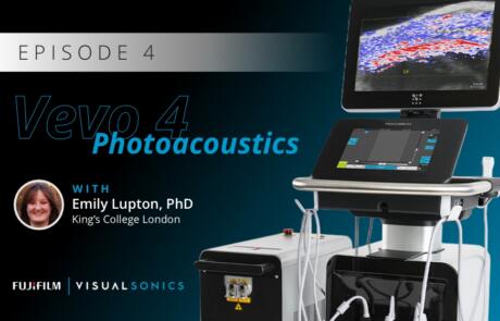
Functional Ultrasound Neuroimaging: Principles, Applications, & Perspectives
Dr. Mickael Tanter presents his work on functional ultrasound neuroimaging, including an overview and translation to clinical applications.
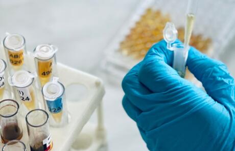
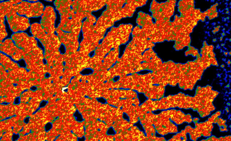
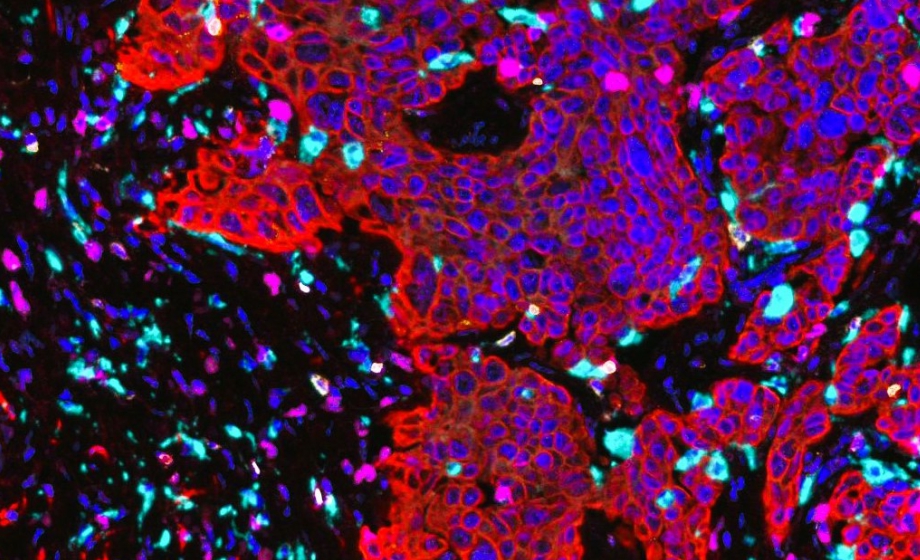
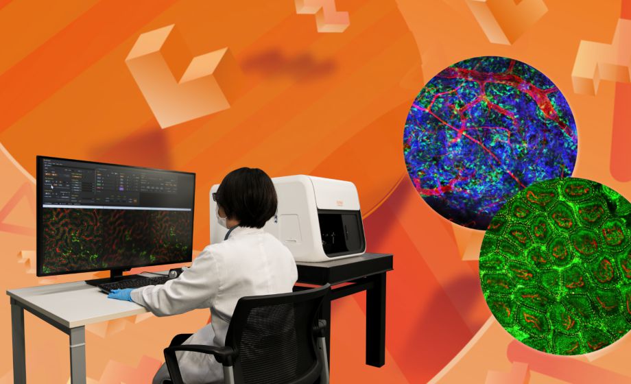
#ExpertAnswers: Lorcan Sherry and Aditya Pratapa on Analyzing Multiplex Immunofluorescence Images
Dr. Lorcan Sherry and Dr. Aditya Pratapa answer questions from a recent webinar where they present a novel workflow for analyzing multiplex immunofluorescence images.
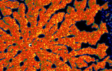
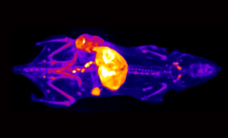
#ExpertAnswers: Matt Silva on Cryo-Fluorescence Tomography for Immunotherapy Research
Matthew Silva, PhD, CEO of EMIT Imaging, answers questions from a recent webinar where he highlights the role of cryo-fluorescence tomography in advancing the field of immunotherapy research.
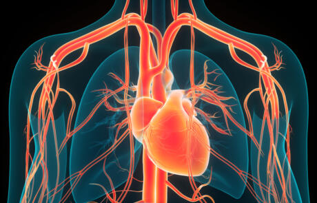
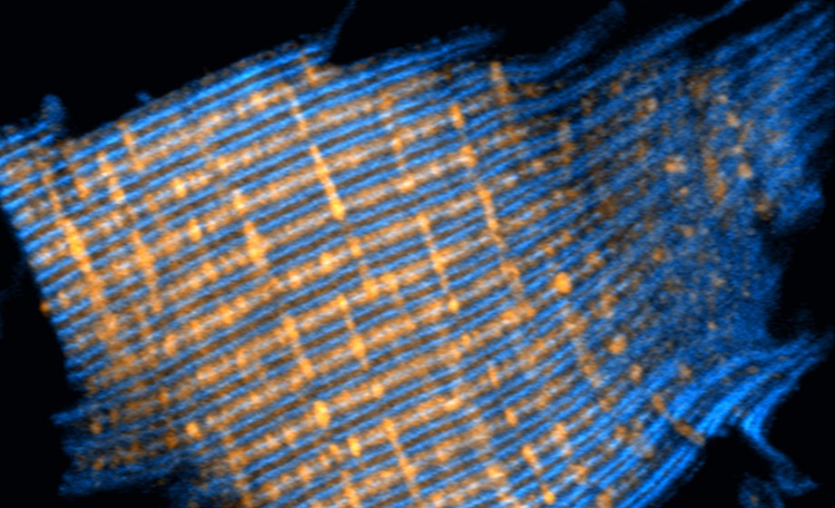
Understanding Advanced Cardiac Tissue Slice Applications
Bradley Palmer, Matthew Caporizzo, and Benjamin Lee answer questions from a recent webinar on how cardiac slices and the IonOptix Cardiac Slice System can be used to complement and improve upon traditional cardiac investigations.
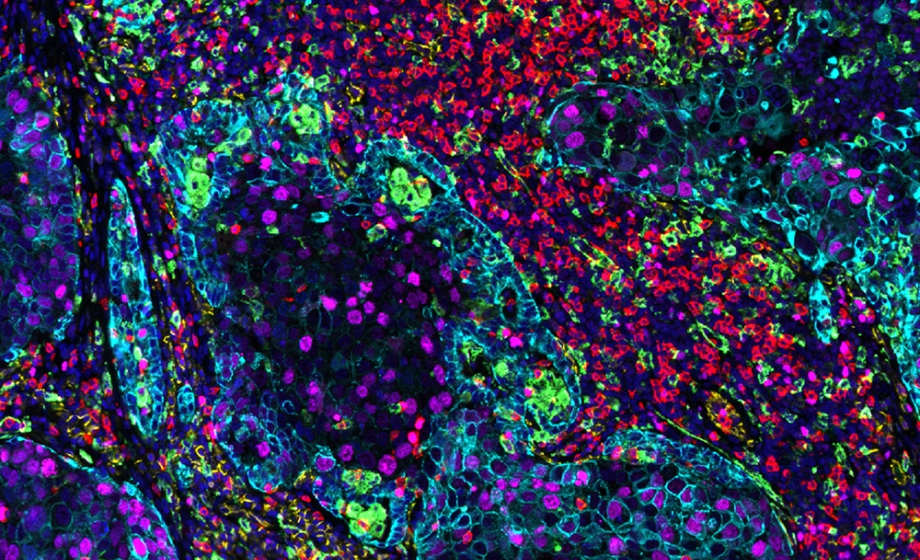
A Ready-to-Analyze High-Plex Spatial Signature Development Workflow for Cancer Immunotherapy
In this webinar, Aditya Pratapa and Lorcan Sherry present a new workflow for analyzing multiplex immunoflurescence images.
Understanding Advanced Cardiac Tissue Slice Applications
Watch to understand how cardiac slices and the IonOptix Cardiac Slice System can be used to complement and improve upon traditional cardiac investigations.
Unlocking Spatial Insights: Accelerate Discoveries 70% Faster with SignalStar™ Multiplex IHC
Join us to learn how to accelerate spatial biology discoveries by 70% with optimized, ready-to-go panels using SignalStar™ Multiplex IHC.
Revolutionizing Immunotherapy Research with 3D, Multiplexed Cryo-fluorescence Tomography
Dr. Matt Silva showcases the power of cryo-fluorescence tomography (CFT) in advancing immunotherapy research.
Real-time IntraVital Microscopy (IVM): In Vivo Live Cell Imaging in Immunology and Neuroscience with Live Imaging Demo
Selina Soyeon Ahn, PhD introduces IVIM Technology’s All-in-One real-time intravital confocal and two-photon microscopy system and its application to immunology and neuroscience research.
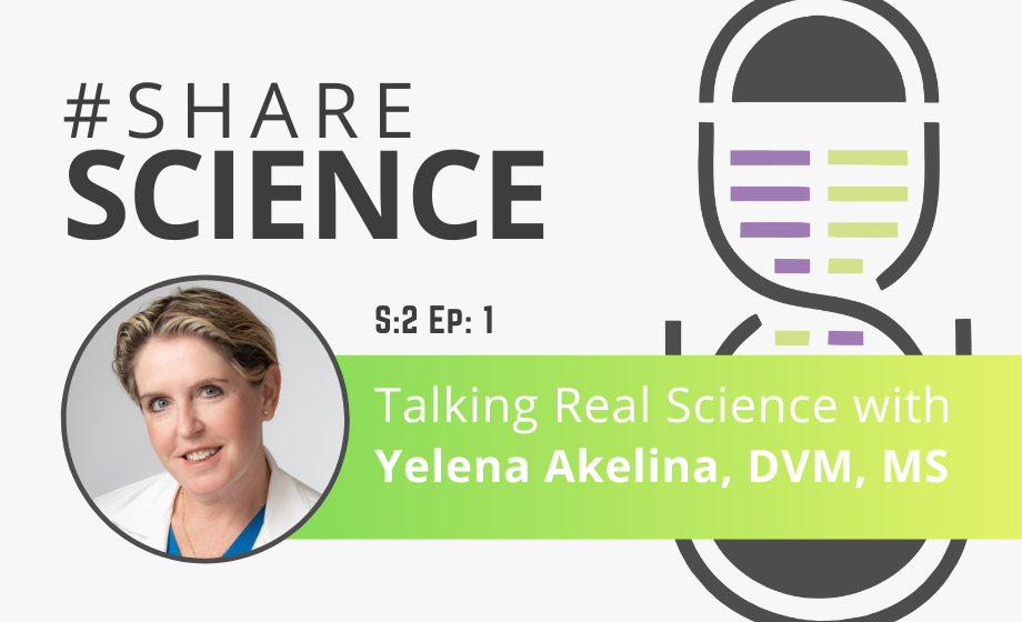
Talking Real Science with Yelena Akelina
This episode of Share Science features Yelena Akelina, DVM, MS, a Research Scientist and a Co-Director/Instructor in Clinical Microsurgery at the Microsurgery Research and Training lab at the Department of Orthopaedic Surgery, Columbia University.
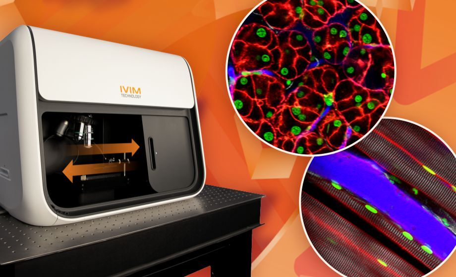
Real-time IntraVital Microscopy (IVM): In Vivo Cellular-level Imaging of Internal Organs in a Live Animal
Join Pilhan Kim, PhD for an in-depth discussion on the All-in-One intravital two-photon and confocal microscopy system.
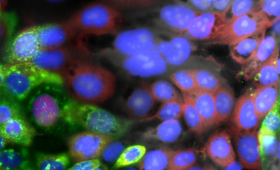
Visualizing the Intracellular Immune Response to SARS-CoV-2 with Fast, High-Content Imaging
Experts discuss how automated microscopy and image analysis can quantify critical markers of infection and describe the insight provided by microscopy in the context of SARS-CoV-2 infection and inflammation.
#ExpertAnswers: Pierre Pouget and Serge Picaud on Functional Ultrasound Imaging
Pierre Pouget and Serge Picaud discuss functional ultrasound imaging and how it can be used to study the brain in behaving non-human primates.
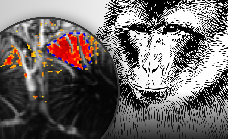
Functional Ultrasound Neuroimaging in Awake & Behaving Non-Human Primates
Dr. Pierre Pouget and Dr. Serge Picaud provide an overview of fUS imaging and discuss how it can be used to study the brain in behaving non-human primates.
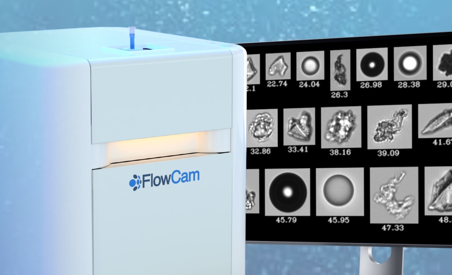
Detection of Protein Aggregation During Biopharmaceutical Development: The Role of Orthogonal Techniques
Dr. Danny Chou discusses the opportunities and challenges in orthogonal particle analysis, and how flow imaging microscopy and light obscuration can be used for formulation development and quality control.
#ExpertAnswers: Alexandra Lubin & Jason Otterstrom on Automated Microscopy
Experts discuss the use of deep learning-powered automated microscopy and image analysis for fast, in vivo zebrafish screening.
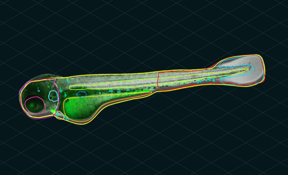
Casting a Wider Net in Zebrafish Screening with Automated Microscopy and Image Analysis
Join Dr. Alexandra Lubin and Dr. Jason Otterstrom as they discuss the use of deep learning-powered automated microscopy and image analysis for fast, in vivo zebrafish screening.