Experts discuss how automated microscopy and image analysis can quantify critical markers of infection and describe the insight provided by microscopy in the context of SARS-CoV-2 infection and inflammation.

To watch this webinar, please contact IDEA Bio-Medical: info@idea-bio.com.

The substantial, real-world impact of SARS-CoV-2 inflammatory disease (COVID-19) is intertwined with the immune response occurring inside of individual cells. The innate immune response is the first line of defense that can stop or slow many types of infection.  It is critical in mounting systemic antiviral responses and signaling to white blood cells (macrophages). SARS-CoV-2 mediated delays of innate immune response activation in airway epithelium cells might provide an opening for the virus to establish itself and shape the outcome of an infection.

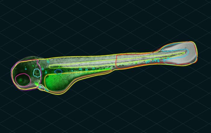
Preservation of the in-situ cellular environment was vital to investigating the interplay between the antiviral and inflammatory roles of the innate immune response to SARS-CoV-2 infection. Automated, high-content imaging (HCI) readily achieves this need and enables direct visualization of the intracellular immune response.

In this webinar, Dr. Jason Otterstrom and Dr. Matthew Whelan describe how automated microscopy and image analysis can quantify critical markers of infection.  Dr. Ann-Kathrin Reuschl, co-lead author of two SARS-CoV-2 infection and inflammation studies, describes the insight provided by microscopy and how it relates to complementary techniques.  Her results reveal that some SARS-CoV-2 variants have evolved the ability to antagonize the intracellular immune response, providing insight into the emergence of subtypes such as the Alpha and Omicron.  Understanding interactions between the virus and the host innate immune response provides a window of opportunity to discover potential therapeutics against severe COVID-19.

Key Topics Include:

  • What is high-content imaging (HCI) and how researchers can use it in 2D cell culture and virology
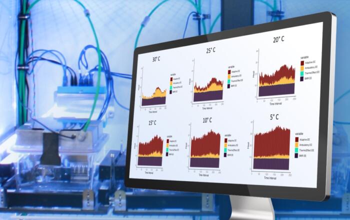
  • How HCI data and insights integrate into larger -omics studies
  • Future directions for HCI in virology to study mechanisms of viral propagation among cells

Who Should Attend?

  • Virologists and immunologists looking to improve their throughput and data quantification using HCI
  • Life-science researchers who want a picture of what is happening in situ with their samples to measure spatial and morphological phenomena
  • People who want to learn how events taking place inside our cells can affect our whole body when we get sick

Resources

To retrieve a PDF copy of the presentation, click on the link below the slide player. From this page, click on the “Download” link to retrieve the file.

Presenters

Research Fellow
HCS Microscopy and Image Analysis
University College London

Dr. Whelan carried out a Bachelor of Science in Cell and Molecular Biology at University College Dublin, focusing on quantitative microscopy of membrane trafficking and perturbation of Golgi morphology in mammalian cells. Upon graduating in 2015, he was awarded the Government of Ireland PhD Research Scholarship (IRC), allowing him to undertake a PhD project of his own design. This student led award afforded him the opportunity to shift his research focus toward investigating the relationship between intracellular microbial pathogens and their hosts. During his PhD, he applied High Content Screening (HCS) microscopy approaches to investigate the effect of DNA supercoiling mediated epigenetic regulation on biofilm formation and host-cell entry by the pathogen Campylobacter jejuni. In 2020, he joined the Division of Infection and Immunity (I&I) in UCL as a Research Fellow in ‘HCS Microscopy and Image Analysis’. Under the supervision of Prof. Madhad Noursadeghi, he developed the ‘Host-Pathogen Microscopy Core’ facility, with the primary goal of providing cutting edge HCS and super-resolution microscopy and quantitation of host cell responses to viral and bacterial infections. He is also currently a postdoctoral researcher in the Laboratory of Prof. Clare Jolly in I&I. His own research focuses on the application of live cell and super-resolution approaches to investigate the kinetics of HIV nuclear entry in primary T cells.

Application Scientist
IDEA Bio-Medical

Dr. Otterstrom is an application scientist for IDEA Bio-Medical. He has a diverse background in biophysics including microscopy, optical design, image analysis and sample labeling. His expertise is adapting biological assays to benefit from utilization of automated microscopy methods.

Postdoctoral Research Associate
Division of Infection & Immunity
University College London

Dr. Ann-Kathrin Reuschl is a postdoctoral Research Associate within the Division of Infection & Immunity at University College London. With a background in infection immunology, she undertook a PhD at Imperial College London during which she explored early interactions between M. tuberculosis and primary human airway epithelium, showing an underappreciated contribution of the epithelium to local inflammation during infection. In 2016, Ann-Kathrin joined Prof Clare Jolly’s team at UCL where she started to interrogate how HIV-1 reprograms its target, the human primary CD4+ T cell. When the pandemic struck, her research pivoted to SARS-CoV-2 host-pathogen interactions. Ann-Kathrin could draw upon her expertise in airway epithelial cell and infection biology to aid the establishment of a SARS-CoV-2 research program at the Division of Infection and Immunity at UCL. As part of a national and international collaboration, she has contributed to fundamental discoveries about SARS-CoV-2 infection biology as well as exploring therapeutic targets. Crucially, this work showed how SARS-CoV-2 evolution allowed the virus to evade the human innate immune response (Thorne et al, Nature, 2021). This work has implications for the molecular host-pathogen interactions of emerging variants that shape the immunological niche in which the virus resides.

Production Partner

IDEA Bio-Medical Ltd.

IDEA Bio-Medical was founded in 2007 through a partnership between YEDA (the Weizmann Institute’s commercialization arm) and IDEA Machine Development (an innovation hub). We specialize in automated imaging systems and image analysis software, offering a broad range of biological applications based on the company’s unique algorithms library. The company is developing novel image-based screening platforms for the pharmaceutical industry and medical centers, dedicated to broadening the scope of personalized medicine. Our WiScan Hermes system incorporates the most advanced technologies currently available in the machine vision field, integrated with engineering methodologies of high reliability and quality at the level of semi-conductors and digital printing industries, which are the specialty of our mother company, IDEA Machine Development Design and Production Ltd.

Additional Content From IDEA Bio-Medical Ltd.

Related Content