Dr. Joanna Hummer and Dr. Florie Le Prieult share insights into cerebral open flow microperfusion (cOFM) use and utility in the broader neuroscience field and specifically for the development of drugs for neurodegenerative diseases.
Highlights
- An introduction to Joanneum Research and cOFM
- cOFM advantages, methods, and applications
- A case study comparing cOFM and microdialysis measurement following amitriptyline administration
- An introduction to biologics and common methods
- An experimental setup for study of biologics using cOFM and microdialysis
- A case study demonstrating the use of cOFM and microdialysis to evaluate target engagement (TE) of anti-Tau antibodies
- A summary of the practical advantages of cOFM and microdialysis
Webinar Summary
This webinar begins with an introduction to the Joanneum Research institute before describing the open flow microperfusion (OFM) technique. This method has been used in ex vivo, preclinical, and clinical studies to examine different targets including dermal, adipose, and brain tissues, and more recently in muscle and mucosa.
“Open flow microperfusion provides unfiltered . . . interstitial fluid [ISF] for unique insights into metabolism and signaling as well as pharmacokinetic and pharmacodynamic studies at the target tissue.”
Dr. Hummer focuses on cOFM and introduces some of the challenges inherent in brain measurements in vivo, such as the presence of the blood brain barrier (BBB), before describing how these can be overcome using microdialysis and cOFM. The method of cOFM sampling is described, including probe implantation, perfusate flow, and sample collection.
“cOFM is a tool to sample cerebral interstitial fluid for the investigation of pharmacokinetics and pharmacodynamics in the brain with intact blood brain barrier.”
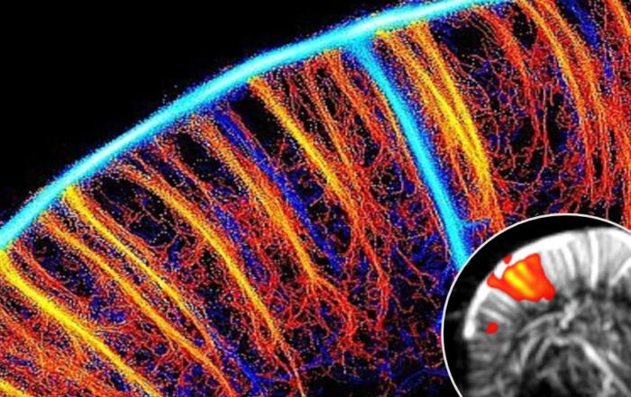
A detailed illustration of cOFM probe implantation is presented with references for scale, followed by use of a guide cannula to preserve BBB integrity before sampling; validation data are also shown illustrating the post-operative recovery of BBB permeability to control levels.
“The cerebral interstitial fluid samples . . . contain all molecules present in the brain tissue without restriction due to size or inherent chemical properties including drugs, proteins, and antibodies, or even larger structures like nanoparticles.”
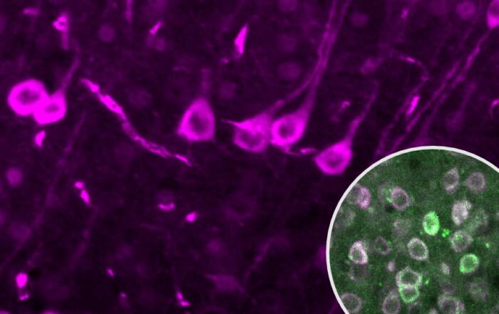
cOFM allows for monitoring substance of interest concentrations in relevant brain regions over time, including development of a complete pharmacokinetic (PK) profile and biomarker responses to intervention for days, or theoretically even months, after implantation. Local tissue reactions to probe implantation are discussed, with astrocyte and macrophage quantification data, demonstrating minimal tissue reaction and the absence of glial scar formation.
“. . . cOFM causes only minimal tissue reaction and the tissue enters a stable condition at 15 days after probe implantation.”
The cOFM probe length and position can be adapted to sample different brain regions and may also be used to sample cerebrospinal fluid; it is also possible to use up to three probes to sample multiple brain regions simultaneously. The potential applications of cOFM are described, and, due to the absence of a probe membrane, a wide range of substances may be monitored regardless of size, lipophilicity, or protein binding properties. This flexibility allows for use of cOFM to monitor BBB changes, PK, and pharmacodynamics (PD) in different species with time-resolved data collection.
A case study is presented in which the antidepressant amitriptyline was injected intraperitoneally and monitored over time by cOFM in one brain hemisphere and compared with microdialysis data collected from the other hemisphere. A number of additional cOFM applications are discussed with accompanying references, including studies of neurodegenerative diseases, obesity and diabetes, inflammation and BBB integrity, nanoparticles, and ongoing work studying glioblastoma. Joanneum Research offers a range of services related to cOFM studies, including probe production and customization, implantation and training, as well as dosing and data collection. In these experiments, the BASi Raturn system is used for sampling in awake and freely moving animals and the Culex system for blood sampling.
“In summary, the full setup allows simultaneous dosing and sampling of cerebral interstitial fluid, cerebrospinal fluid, and blood in awake and freely moving animals over several days.”
Dr. Hummer concludes her portion of the webinar with a summary of the features, advantages, and opportunities made possible using cOFM.
Dr. Le Prieult begins the second half of the webinar with an introduction to biologics, including the brain to plasma or serum ratio, the challenge of brain penetration, and typical methodologies employed. A summary of the experimental setup used is shown, including the Raturn system introduced previously, and their workflow for measuring antibody concentrations. Quality controls are considered, including flow monitoring, tissue pathology at probe insertion sites, residual blood minimization in tissue samples, and BBB integrity assessment. Data collected in mice following intraperitoneal or intravenous administration of a non-target binding antibody are shown, together with measurement of PK parameters. Dr. Le Prieult proceeds to describe the approach used to obtain absolute quantification of cOFM data, corrected for dilution introduced by the push-pull perfusion system, and achieved by introducing step-wise changes in flow rate. cOFM allows for data collection over long time periods in a single animal, including extended recording immediately after antibody administration and again one week later.
Dr. Le Prieult starts the final portion of her presentation with a case study demonstrating the use of microdialysis to study TE of anti-Tau monoclonal antibodies; this would not be possible using standard methods such as brain homogenate assays. The results clearly demonstrate effective TE following high affinity anti-Tau antibody administration as reflected in a decrease of free Tau in the interstitial fluid.
“. . . through this tool and the implementation of this highly sensitive measuring method, we could actually have both . . . PK and PD in the same samples . . .”
In conclusion, Dr. Le Prieult summarizes the benefits and opportunities of cOFM and microdialysis methods, including antibody and biomarker quantification in brain ISF, detailed time resolution of PK data, high sensitivity TE assessment in parallel with other analytical approaches, and provision of data inputs for clinically-relevant models of expected ISF and CSF antibody concentrations.
Click to watch the webinar recording. To view the presentation full screen simply click the square icon located in the bottom-right corner of the video-viewer.
Resources


Q&A
- What are the considerations for use of cOFM in larger species?
- Is purification of the sample required for cOFM?
- Is the age of the animals important, and does this affect the recovery period?
- Has this been used in neonatal rodents or in the study of traumatic brain injury models?
- What is the area coverage of the cOFM method?
- Why were recovery-corrected microdialysis data compared with non-recovery-corrected cOFM data?
- What flow rates were used?
- What is the longest possible sampling period?
- Has this method been used in aged animals, and does it cause additional trauma in comparison with younger animals?
- Can animals be sent to Joanneum Research for study?
- Can cOFM be used to deliver substances to the brain?
- Do you have any data on the stability of collected cOFM samples?
To retrieve a PDF copy of the presentation, click on the link below the slide player. From this page, click on the “Download” link to retrieve the file.
Presenters
Deputy Head of Biomedical Tissue Monitoring
HEALTH - Institute for Biomedicine and Health Sciences
JOANNEUM RESEARCH Forschungsgesellschaft m.b.H
Lab Head
In vivo Neuroscience Pharmacokinetics
AbbVie
Production Partner
BASi – Bioanalytical Systems, Inc.

JOANNEUM RESEARCH HEALTH